5OAK

Structure of the dmPar3 PDZ1 domain in complex with the dmPar6 PBM


Experimental Data Snapshot

  • Method: X-RAY DIFFRACTION
  • Resolution: 1.50 Å
  • R-Value Free: 0.169 
  • R-Value Work: 0.140 
  • R-Value Observed: 0.142 

wwPDB Validation   3D Report Full Report


This is version 1.2 of the entry. See complete history


Literature

Structural basis for the interaction between the cell polarity proteins Par3 and Par6.

Renschler, F.A.Bruekner, S.R.Salomon, P.L.Mukherjee, A.Kullmann, L.Schutz-Stoffregen, M.C.Henzler, C.Pawson, T.Krahn, M.P.Wiesner, S.

(2018) Sci Signal 11

  • DOI: https://doi.org/10.1126/scisignal.aam9899
  • Primary Citation of Related Structures:  
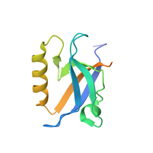
    5OAK

  • PubMed Abstract: 

    Polarity is a fundamental property of most cell types. The Par protein complex is a major driving force in generating asymmetrically localized protein networks and consists of atypical protein kinase C (aPKC), Par3, and Par6. Dysfunction of this complex causes developmental abnormalities and diseases such as cancer. We identified a PDZ domain-binding motif in Par6 that was essential for its interaction with Par3 in vitro and for Par3-mediated membrane localization of Par6 in cultured cells. In fly embryos, we observed that the PDZ domain-binding motif was functionally redundant with the PDZ domain in targeting Par6 to the cortex of epithelial cells. Our structural analyses by x-ray crystallography and NMR spectroscopy showed that both the PDZ1 and PDZ3 domains but not the PDZ2 domain in Par3 engaged in a canonical interaction with the PDZ domain-binding motif in Par6. Par3 thus has the potential to recruit two Par6 proteins simultaneously, which may facilitate the assembly of polarity protein networks through multivalent PDZ domain interactions.


  • Organizational Affiliation

    Max Planck Institute for Developmental Biology, Max-Planck-Ring 5, 72076 Tübingen, Germany.


Macromolecules
Find similar proteins by:  (by identity cutoff)  |  3D Structure
Entity ID: 1
MoleculeChains Sequence LengthOrganismDetailsImage
Bazooka, isoform C,LD29223p
A, B, C, D
117Drosophila melanogasterMutation(s): 0 
Gene Names: bazCG5055Dmel_CG5055par-6CG5884Dmel_CG5884
UniProt
Find proteins for Q0KHR3 (Drosophila melanogaster)
Explore Q0KHR3 
Go to UniProtKB:  Q0KHR3
Entity Groups  
Sequence Clusters30% Identity50% Identity70% Identity90% Identity95% Identity100% Identity
UniProt GroupQ0KHR3
Sequence Annotations
Expand
  • Reference Sequence
Small Molecules
Ligands 1 Unique
IDChains Name / Formula / InChI Key2D Diagram3D Interactions
GOL
Query on GOL

Download Ideal Coordinates CCD File 
E [auth A]GLYCEROL
C3 H8 O3
PEDCQBHIVMGVHV-UHFFFAOYSA-N
Experimental Data & Validation

Experimental Data

  • Method: X-RAY DIFFRACTION
  • Resolution: 1.50 Å
  • R-Value Free: 0.169 
  • R-Value Work: 0.140 
  • R-Value Observed: 0.142 
  • Space Group: P 31
Unit Cell:
Length ( Å )Angle ( ˚ )
a = 60.88α = 90
b = 60.88β = 90
c = 65.05γ = 120
Software Package:
Software NamePurpose
XSCALEdata scaling
PHENIXrefinement
PDB_EXTRACTdata extraction
XDSdata reduction
PHASERphasing

Structure Validation

View Full Validation Report


Entry History & Funding Information

Deposition Data


Funding OrganizationLocationGrant Number
Max-Planck GesellschaftGermany--
IMPRS "From molecules to Organisms"Germany--

Revision History  (Full details and data files)

  • Version 1.0: 2018-02-21
    Type: Initial release
  • Version 1.1: 2018-02-28
    Changes: Database references
  • Version 1.2: 2024-01-17
    Changes: Data collection, Database references, Refinement description