5O99

Unconventional SH3 domain from the postsynaptic density scaffold protein Shank3


Experimental Data Snapshot

  • Method: X-RAY DIFFRACTION
  • Resolution: 0.87 Å
  • R-Value Free: 0.117 
  • R-Value Work: 0.097 
  • R-Value Observed: 0.112 

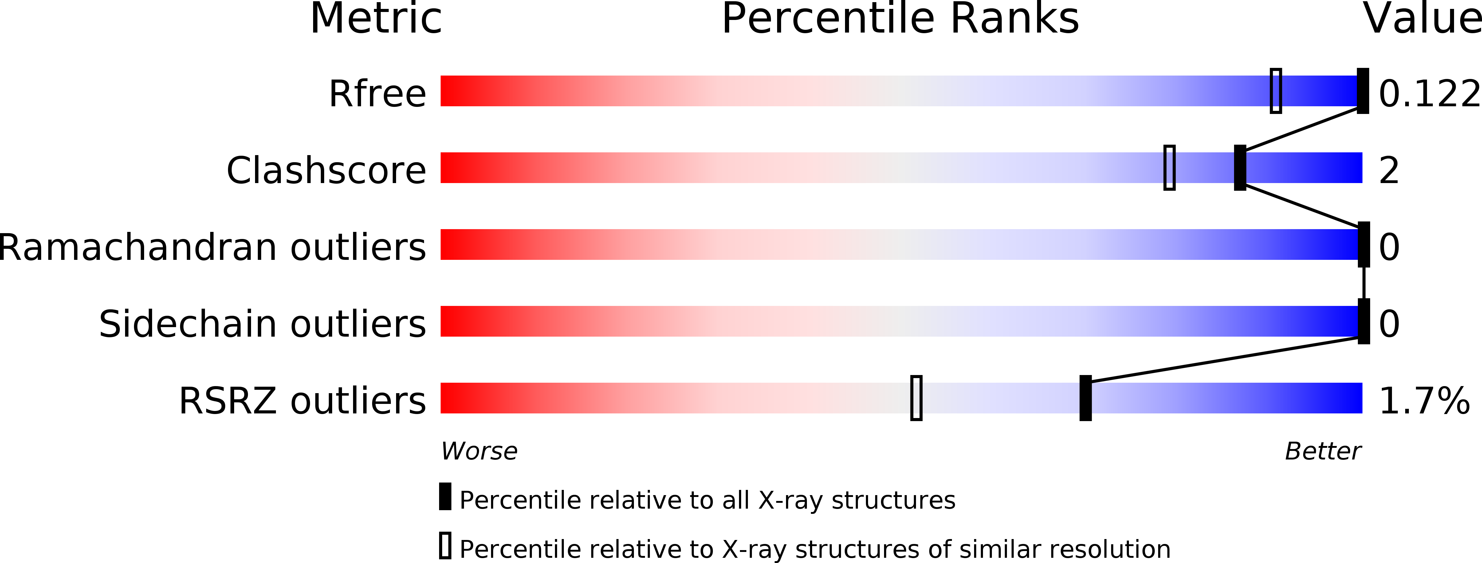
wwPDB Validation   3D Report Full Report


This is version 1.1 of the entry. See complete history


Literature

Structure of an unconventional SH3 domain from the postsynaptic density protein Shank3 at ultrahigh resolution.

Ponna, S.K.Myllykoski, M.Boeckers, T.M.Kursula, P.

(2017) Biochem Biophys Res Commun 490: 806-812

  • DOI: https://doi.org/10.1016/j.bbrc.2017.06.121
  • Primary Citation of Related Structures:  
    5O99

  • PubMed Abstract: 

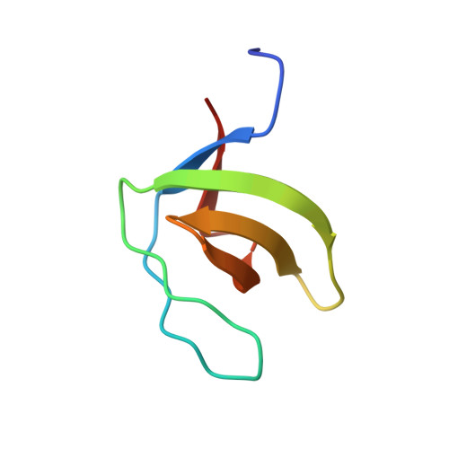
    The Shank family comprises three large multi-domain proteins playing central roles as protein scaffolds in the neuronal postsynaptic density. The Shank proteins are closely linked to neuropsychiatric diseases, such as autism spectrum disorders. One characteristic domain in the Shank family is the SH3 domain, assumed to play a role in protein-protein interactions; however, no specific ligand binding to any Shank SH3 domain has been described. We solved the crystal structure of the SH3 domain from Shank3 at sub-atomic resolution. While the structure presents the canonical SH3 domain fold, the binding site for proline-rich peptides is not conserved. In line with this, no binding of Pro-rich sequences by the Shank3 SH3 domain was observed. Sequence comparisons indicate that all Shank isoforms have similarly lost the classical Pro-rich peptide binding site from the SH3 domain. Whether the corresponding site in the Shank SH3 domains has evolved to bind a non-poly-Pro target sequence is currently not known. Our work provides an intriguing example of the evolution of a well-characterized protein-protein interaction domain within the context of multi-domain protein scaffolds, allowing the conservation of structural features, but losing canonical functional sites. The data are further discussed in light of known mutations in the SH3 domain or its vicinity in the different Shank isoforms.


  • Organizational Affiliation

    Faculty of Biochemistry and Molecular Medicine & Biocenter Oulu, University of Oulu, Finland.


Macromolecules
Find similar proteins by:  (by identity cutoff)  |  3D Structure
Entity ID: 1
MoleculeChains Sequence LengthOrganismDetailsImage
SH3 and multiple ankyrin repeat domains protein 3A [auth B],
B [auth A]
60Rattus norvegicusMutation(s): 0 
Gene Names: Shank3Prosap2
UniProt
Find proteins for Q9JLU4 (Rattus norvegicus)
Explore Q9JLU4 
Go to UniProtKB:  Q9JLU4
Entity Groups  
Sequence Clusters30% Identity50% Identity70% Identity90% Identity95% Identity100% Identity
UniProt GroupQ9JLU4
Sequence Annotations
Expand
  • Reference Sequence
Small Molecules
Ligands 1 Unique
IDChains Name / Formula / InChI Key2D Diagram3D Interactions
BTB
Query on BTB

Download Ideal Coordinates CCD File 
C [auth A]2-[BIS-(2-HYDROXY-ETHYL)-AMINO]-2-HYDROXYMETHYL-PROPANE-1,3-DIOL
C8 H19 N O5
OWMVSZAMULFTJU-UHFFFAOYSA-N
Experimental Data & Validation

Experimental Data

  • Method: X-RAY DIFFRACTION
  • Resolution: 0.87 Å
  • R-Value Free: 0.117 
  • R-Value Work: 0.097 
  • R-Value Observed: 0.112 
  • Space Group: P 1 21 1
Unit Cell:
Length ( Å )Angle ( ˚ )
a = 29.74α = 90
b = 52.91β = 90.05
c = 31.66γ = 90
Software Package:
Software NamePurpose
PHENIXrefinement
XDSdata reduction
XDSdata scaling
Auto-Rickshawphasing

Structure Validation

View Full Validation Report


Entry History & Funding Information

Deposition Data


Funding OrganizationLocationGrant Number
Sigrid Juselius FoundationFinland--
Emil Aaltonen FoundationFinland--

Revision History  (Full details and data files)

  • Version 1.0: 2017-07-05
    Type: Initial release
  • Version 1.1: 2017-08-02
    Changes: Database references