5O90

Crystal structure of a P38alpha T185G mutant in complex with TAB1 peptide.


Experimental Data Snapshot

  • Method: X-RAY DIFFRACTION
  • Resolution: 2.49 Å
  • R-Value Free: 0.275 
  • R-Value Work: 0.249 
  • R-Value Observed: 0.250 

wwPDB Validation   3D Report Full Report


Ligand Structure Quality Assessment 


This is version 1.2 of the entry. See complete history


Literature

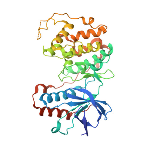
TAB1-Induced Autoactivation of p38 alpha Mitogen-Activated Protein Kinase Is Crucially Dependent on Threonine 185.

Thapa, D.Nichols, C.Bassi, R.Martin, E.D.Verma, S.Conte, M.R.De Santis, V.De Nicola, G.F.Marber, M.S.

(2018) Mol Cell Biol 38

  • DOI: https://doi.org/10.1128/MCB.00409-17
  • Primary Citation of Related Structures:  
    5O90

  • PubMed Abstract: 

    p38α mitogen-activated protein kinase is essential to cellular homeostasis. Two principal mechanisms to activate p38α exist. The first relies on dedicated dual-specificity kinases such as mitogen-activated protein kinase kinase (MAP2K) 3 (MKK3) or 6 (MKK6), which activate p38α by phosphorylating Thr180 and Tyr182 within the activation segment. The second is by autophosphorylation of Thr180 and Tyr182 in cis , mediated by p38α binding the scaffold protein TAB1. The second mechanism occurs during myocardial ischemia, where it aggravates myocardial infarction. Based on the crystal structure of the p38α-TAB1 complex we replaced threonine 185 of p38α with glycine (T185G) to prevent an intramolecular hydrogen bond with Asp150 from being formed. This mutation did not interfere with TAB1 binding to p38α. However, it disrupted the consequent long-range effect of this binding event on the distal activation segment, releasing the constraint on Thr180 that oriented its hydroxyl for phosphotransfer. Based on assays performed in vitro and in vivo , the autoactivation of p38α(T185G) was disabled, while its ability to be activated by upstream MAP2Ks and to phosphorylate downstream substrates remained intact. Furthermore, myocardial cells expressing p38α(T185G) were resistant to injury. These findings reveal a mechanism to selectively disable p38α autoactivation and its consequences, which may ultimately circumvent the toxicity associated with strategies that inhibit p38α kinase activity under all circumstances, such as with ATP-competitive inhibitors.


  • Organizational Affiliation

    British Heart Foundation Centre of Excellence, Department of Cardiology, The Rayne Institute, St Thomas' Hospital, King's College London, London, United Kingdom.


Macromolecules
Find similar proteins by:  (by identity cutoff)  |  3D Structure
Entity ID: 1
MoleculeChains Sequence LengthOrganismDetailsImage
Mitogen-activated protein kinase 14360Mus musculusMutation(s): 1 
Gene Names: Mapk14Crk1Csbp1Csbp2
EC: 2.7.11.24
UniProt
Find proteins for P47811 (Mus musculus)
Explore P47811 
Go to UniProtKB:  P47811
Entity Groups  
Sequence Clusters30% Identity50% Identity70% Identity90% Identity95% Identity100% Identity
UniProt GroupP47811
Sequence Annotations
Expand
  • Reference Sequence
Find similar proteins by:  (by identity cutoff)  |  3D Structure
Entity ID: 2
MoleculeChains Sequence LengthOrganismDetailsImage
TGF-beta-activated kinase 1 and MAP3K7-binding protein 129Homo sapiensMutation(s): 0 
UniProt & NIH Common Fund Data Resources
Find proteins for Q15750 (Homo sapiens)
Explore Q15750 
Go to UniProtKB:  Q15750
PHAROS:  Q15750
GTEx:  ENSG00000100324 
Entity Groups  
Sequence Clusters30% Identity50% Identity70% Identity90% Identity95% Identity100% Identity
UniProt GroupQ15750
Sequence Annotations
Expand
  • Reference Sequence
Small Molecules
Ligands 1 Unique
IDChains Name / Formula / InChI Key2D Diagram3D Interactions
SB4
Query on SB4

Download Ideal Coordinates CCD File 
C [auth A]4-(4-FLUOROPHENYL)-1-(4-PIPERIDINYL)-5-(2-AMINO-4-PYRIMIDINYL)-IMIDAZOLE
C18 H19 F N6
VSPFURGQAYMVAN-UHFFFAOYSA-N
Binding Affinity Annotations 
IDSourceBinding Affinity
SB4 BindingDB:  5O90 IC50: min: 19, max: 20 (nM) from 2 assay(s)
Experimental Data & Validation

Experimental Data

  • Method: X-RAY DIFFRACTION
  • Resolution: 2.49 Å
  • R-Value Free: 0.275 
  • R-Value Work: 0.249 
  • R-Value Observed: 0.250 
  • Space Group: P 1 21 1
Unit Cell:
Length ( Å )Angle ( ˚ )
a = 45.92α = 90
b = 73.58β = 91.09
c = 58.97γ = 90
Software Package:
Software NamePurpose
PHENIXrefinement
xia2data reduction
xia2data scaling
PHASERphasing

Structure Validation

View Full Validation Report


Ligand Structure Quality Assessment 


Entry History & Funding Information

Deposition Data


Funding OrganizationLocationGrant Number
BHFUnited KingdomBHF - FS/14/29/30896 - DE NICOLA

Revision History  (Full details and data files)

  • Version 1.0: 2018-01-31
    Type: Initial release
  • Version 1.1: 2018-02-21
    Changes: Database references
  • Version 1.2: 2024-01-17
    Changes: Data collection, Database references, Refinement description