5O5L

X-ray structure of a bacterial adenylyl cyclase soluble domain, solved at cryogenic temperature


Experimental Data Snapshot

  • Method: X-RAY DIFFRACTION
  • Resolution: 2.70 Å
  • R-Value Free: 0.289 
  • R-Value Work: 0.265 
  • R-Value Observed: 0.267 

wwPDB Validation   3D Report Full Report


Ligand Structure Quality Assessment 


This is version 1.3 of the entry. See complete history


Literature

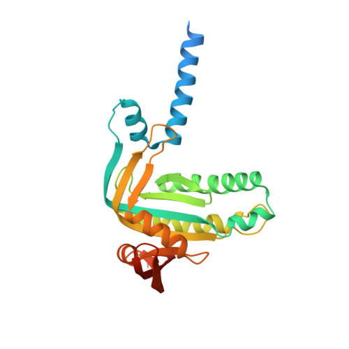
Role of the nucleotidyl cyclase helical domain in catalytically active dimer formation.

Vercellino, I.Rezabkova, L.Olieric, V.Polyhach, Y.Weinert, T.Kammerer, R.A.Jeschke, G.Korkhov, V.M.

(2017) Proc Natl Acad Sci U S A 114: E9821-E9828

  • DOI: https://doi.org/10.1073/pnas.1712621114
  • Primary Citation of Related Structures:  
    5O5K, 5O5L

  • PubMed Abstract: 

    Nucleotidyl cyclases, including membrane-integral and soluble adenylyl and guanylyl cyclases, are central components in a wide range of signaling pathways. These proteins are architecturally diverse, yet many of them share a conserved feature, a helical region that precedes the catalytic cyclase domain. The role of this region in cyclase dimerization has been a subject of debate. Although mutations within this region in various cyclases have been linked to genetic diseases, the molecular details of their effects on the enzymes remain unknown. Here, we report an X-ray structure of the cytosolic portion of the membrane-integral adenylyl cyclase Cya from Mycobacterium intracellulare in a nucleotide-bound state. The helical domains of each Cya monomer form a tight hairpin, bringing the two catalytic domains into an active dimerized state. Mutations in the helical domain of Cya mimic the disease-related mutations in human proteins, recapitulating the profiles of the corresponding mutated enzymes, adenylyl cyclase-5 and retinal guanylyl cyclase-1. Our experiments with full-length Cya and its cytosolic domain link the mutations to protein stability, and the ability to induce an active dimeric conformation of the catalytic domains. Sequence conservation indicates that this domain is an integral part of cyclase machinery across protein families and species. Our study provides evidence for a role of the helical domain in establishing a catalytically competent dimeric cyclase conformation. Our results also suggest that the disease-associated mutations in the corresponding regions of human nucleotidyl cyclases disrupt the normal helical domain structure.


  • Organizational Affiliation

    Laboratory of Biomolecular Research, Division of Biology and Chemistry, Paul Scherrer Institute, 5232 Villigen, Switzerland.


Macromolecules
Find similar proteins by:  (by identity cutoff)  |  3D Structure
Entity ID: 1
MoleculeChains Sequence LengthOrganismDetailsImage
Adenylate cyclase
A, B, C, D, E
A, B, C, D, E, F, G, H, I, J, K
254Mycobacterium intracellulare 1956Mutation(s): 0 
Gene Names: cyaI550_3099
EC: 4.6.1.1
UniProt
Find proteins for X8CHM4 (Mycobacterium intracellulare 1956)
Explore X8CHM4 
Go to UniProtKB:  X8CHM4
Entity Groups  
Sequence Clusters30% Identity50% Identity70% Identity90% Identity95% Identity100% Identity
UniProt GroupX8CHM4
Sequence Annotations
Expand
  • Reference Sequence
Small Molecules
Ligands 3 Unique
IDChains Name / Formula / InChI Key2D Diagram3D Interactions
ONM
Query on ONM

Download Ideal Coordinates CCD File 
CA [auth E]
GA [auth F]
L [auth A]
LA [auth G]
MA [auth H]
CA [auth E],
GA [auth F],
L [auth A],
LA [auth G],
MA [auth H],
Q [auth B],
RA [auth I],
SA [auth J],
U [auth C],
VA [auth K],
Z [auth D]
3'-O-(N-METHYLANTHRANILOYL)-GUANOSINE-5'-TRIPHOSPHATE
C18 H23 N6 O15 P3
DSPRYHPLXXUNHS-XNIJJKJLSA-N
SO4
Query on SO4

Download Ideal Coordinates CCD File 
DA [auth E]
O [auth A]
P [auth B]
R [auth B]
V [auth C]
DA [auth E],
O [auth A],
P [auth B],
R [auth B],
V [auth C],
Y [auth D]
SULFATE ION
O4 S
QAOWNCQODCNURD-UHFFFAOYSA-L
MN
Query on MN

Download Ideal Coordinates CCD File 
AA [auth D]
BA [auth D]
EA [auth E]
FA [auth E]
HA [auth F]
AA [auth D],
BA [auth D],
EA [auth E],
FA [auth E],
HA [auth F],
IA [auth F],
JA [auth G],
KA [auth G],
M [auth A],
N [auth A],
NA [auth H],
OA [auth H],
PA [auth I],
QA [auth I],
S [auth B],
T [auth B],
TA [auth J],
UA [auth J],
W [auth C],
WA [auth K],
X [auth C],
XA [auth K]
MANGANESE (II) ION
Mn
WAEMQWOKJMHJLA-UHFFFAOYSA-N
Experimental Data & Validation

Experimental Data

  • Method: X-RAY DIFFRACTION
  • Resolution: 2.70 Å
  • R-Value Free: 0.289 
  • R-Value Work: 0.265 
  • R-Value Observed: 0.267 
  • Space Group: C 1 2 1
Unit Cell:
Length ( Å )Angle ( ˚ )
a = 144.48α = 90
b = 84.12β = 103.87
c = 310.75γ = 90
Software Package:
Software NamePurpose
BUSTERrefinement
XDSdata reduction
XSCALEdata scaling
PHENIXphasing

Structure Validation

View Full Validation Report


Ligand Structure Quality Assessment 


Entry History & Funding Information

Deposition Data


Funding OrganizationLocationGrant Number
Swiss National Science FoundationSwitzerland150665

Revision History  (Full details and data files)

  • Version 1.0: 2017-11-01
    Type: Initial release
  • Version 1.1: 2017-11-08
    Changes: Database references
  • Version 1.2: 2017-11-22
    Changes: Database references
  • Version 1.3: 2019-10-16
    Changes: Data collection