


Crystal structure of WNK3 kinase and CCT1 didomain in a unphosphorylated state
Pinkas, D.M., Daubner, G.M., Bufton, J.C., Bartual, S.G., Sanvitale, C.E., Alessi, D.R., Bullock, A.To be published.
Experimental Data Snapshot
wwPDB Validation 3D Report Full Report

Entity ID: 1 | |||||
|---|---|---|---|---|---|
| Molecule | Chains | Sequence Length | Organism | Details | Image |
| Serine/threonine-protein kinase WNK3 | 380 | Homo sapiens | Mutation(s): 0 Gene Names: WNK3, KIAA1566, PRKWNK3 EC: 2.7.11.1 | 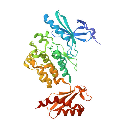 | |
UniProt & NIH Common Fund Data Resources | |||||
Find proteins for Q9BYP7 (Homo sapiens) Explore Q9BYP7 Go to UniProtKB: Q9BYP7 | |||||
PHAROS: Q9BYP7 GTEx: ENSG00000196632 | |||||
Entity Groups | |||||
| Sequence Clusters | 30% Identity50% Identity70% Identity90% Identity95% Identity100% Identity | ||||
| UniProt Group | Q9BYP7 | ||||
Sequence AnnotationsExpand | |||||
| |||||
| Ligands 2 Unique | |||||
|---|---|---|---|---|---|
| ID | Chains | Name / Formula / InChI Key | 2D Diagram | 3D Interactions | |
| PEG Query on PEG | B [auth A] | DI(HYDROXYETHYL)ETHER C4 H10 O3 MTHSVFCYNBDYFN-UHFFFAOYSA-N |  | ||
| GOL Query on GOL | C [auth A], D [auth A], E [auth A], F [auth A] | GLYCEROL C3 H8 O3 PEDCQBHIVMGVHV-UHFFFAOYSA-N |  | ||
| Length ( Å ) | Angle ( ˚ ) |
|---|---|
| a = 166.37 | α = 90 |
| b = 64.08 | β = 97.16 |
| c = 47.34 | γ = 90 |
| Software Name | Purpose |
|---|---|
| BUSTER | refinement |
| XDS | data reduction |
| XSCALE | data scaling |
| PHASER | phasing |
RCSB PDB (citation) is hosted by
RCSB PDB is a member of the

