5O0X

Crystal structure of dehydrogenase domain of Cylindrospermum stagnale NADPH-Oxidase 5 (NOX5)


Experimental Data Snapshot

  • Method: X-RAY DIFFRACTION
  • Resolution: 2.20 Å
  • R-Value Free: 0.209 
  • R-Value Work: 0.184 
  • R-Value Observed: 0.185 

wwPDB Validation   3D Report Full Report


Ligand Structure Quality Assessment 


This is version 1.1 of the entry. See complete history


Literature

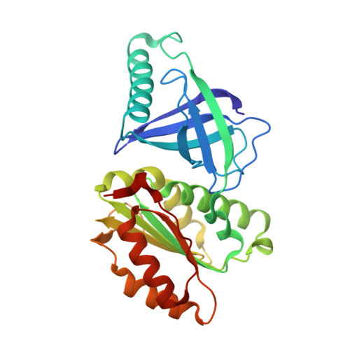
Crystal structures and atomic model of NADPH oxidase.

Magnani, F.Nenci, S.Millana Fananas, E.Ceccon, M.Romero, E.Fraaije, M.W.Mattevi, A.

(2017) Proc Natl Acad Sci U S A 114: 6764-6769

  • DOI: https://doi.org/10.1073/pnas.1702293114
  • Primary Citation of Related Structures:  
    5O0T, 5O0X

  • PubMed Abstract: 

    NADPH oxidases (NOXs) are the only enzymes exclusively dedicated to reactive oxygen species (ROS) generation. Dysregulation of these polytopic membrane proteins impacts the redox signaling cascades that control cell proliferation and death. We describe the atomic crystal structures of the catalytic flavin adenine dinucleotide (FAD)- and heme-binding domains of Cylindrospermum stagnale NOX5. The two domains form the core subunit that is common to all seven members of the NOX family. The domain structures were then docked in silico to provide a generic model for the NOX family. A linear arrangement of cofactors (NADPH, FAD, and two membrane-embedded heme moieties) injects electrons from the intracellular side across the membrane to a specific oxygen-binding cavity on the extracytoplasmic side. The overall spatial organization of critical interactions is revealed between the intracellular loops on the transmembrane domain and the NADPH-oxidizing dehydrogenase domain. In particular, the C terminus functions as a toggle switch, which affects access of the NADPH substrate to the enzyme. The essence of this mechanistic model is that the regulatory cues conformationally gate NADPH-binding, implicitly providing a handle for activating/deactivating the very first step in the redox chain. Such insight provides a framework to the discovery of much needed drugs that selectively target the distinct members of the NOX family and interfere with ROS signaling.


  • Organizational Affiliation

    Department of Biology and Biotechnology "L. Spallanzani," University of Pavia, 27100 Pavia, Italy; francesca.magnani@unipv.it andrea.mattevi@unipv.it.


Macromolecules
Find similar proteins by:  (by identity cutoff)  |  3D Structure
Entity ID: 1
MoleculeChains Sequence LengthOrganismDetailsImage
Putative ferric reductase291Cylindrospermum stagnale PCC 7417Mutation(s): 0 
Gene Names: Cylst_1289
UniProt
Find proteins for K9WT99 (Cylindrospermum stagnale PCC 7417)
Explore K9WT99 
Go to UniProtKB:  K9WT99
Entity Groups  
Sequence Clusters30% Identity50% Identity70% Identity90% Identity95% Identity100% Identity
UniProt GroupK9WT99
Sequence Annotations
Expand
  • Reference Sequence
Small Molecules
Ligands 5 Unique
IDChains Name / Formula / InChI Key2D Diagram3D Interactions
FAD
Query on FAD

Download Ideal Coordinates CCD File 
B [auth A]FLAVIN-ADENINE DINUCLEOTIDE
C27 H33 N9 O15 P2
VWWQXMAJTJZDQX-UYBVJOGSSA-N
P6G
Query on P6G

Download Ideal Coordinates CCD File 
J [auth A],
K [auth A],
L [auth A],
M [auth A],
N [auth A]
HEXAETHYLENE GLYCOL
C12 H26 O7
IIRDTKBZINWQAW-UHFFFAOYSA-N
PEG
Query on PEG

Download Ideal Coordinates CCD File 
CA [auth A]
O [auth A]
P [auth A]
Q [auth A]
R [auth A]
CA [auth A],
O [auth A],
P [auth A],
Q [auth A],
R [auth A],
S [auth A],
T [auth A],
U [auth A],
V [auth A]
DI(HYDROXYETHYL)ETHER
C4 H10 O3
MTHSVFCYNBDYFN-UHFFFAOYSA-N
GOL
Query on GOL

Download Ideal Coordinates CCD File 
AA [auth A]
BA [auth A]
W [auth A]
X [auth A]
Y [auth A]
AA [auth A],
BA [auth A],
W [auth A],
X [auth A],
Y [auth A],
Z [auth A]
GLYCEROL
C3 H8 O3
PEDCQBHIVMGVHV-UHFFFAOYSA-N
CL
Query on CL

Download Ideal Coordinates CCD File 
C [auth A]
D [auth A]
E [auth A]
F [auth A]
G [auth A]
C [auth A],
D [auth A],
E [auth A],
F [auth A],
G [auth A],
H [auth A],
I [auth A]
CHLORIDE ION
Cl
VEXZGXHMUGYJMC-UHFFFAOYSA-M
Experimental Data & Validation

Experimental Data

  • Method: X-RAY DIFFRACTION
  • Resolution: 2.20 Å
  • R-Value Free: 0.209 
  • R-Value Work: 0.184 
  • R-Value Observed: 0.185 
  • Space Group: P 3 2 1
Unit Cell:
Length ( Å )Angle ( ˚ )
a = 128.279α = 90
b = 128.279β = 90
c = 72.519γ = 120
Software Package:
Software NamePurpose
REFMACrefinement
XDSdata reduction
Aimlessdata scaling
PHASERphasing

Structure Validation

View Full Validation Report


Ligand Structure Quality Assessment 


Entry History & Funding Information

Deposition Data


Funding OrganizationLocationGrant Number
Italian Ministry of Science and EducationItalyPRIN2015-20152TE5PK_00

Revision History  (Full details and data files)

  • Version 1.0: 2017-06-28
    Type: Initial release
  • Version 1.1: 2017-07-05
    Changes: Database references