5NUV

Structure of the WD40-domain of human ATG16L1


Experimental Data Snapshot

  • Method: X-RAY DIFFRACTION
  • Resolution: 1.55 Å
  • R-Value Free: 0.197 
  • R-Value Work: 0.165 
  • R-Value Observed: 0.167 

wwPDB Validation   3D Report Full Report


This is version 1.3 of the entry. See complete history


Literature

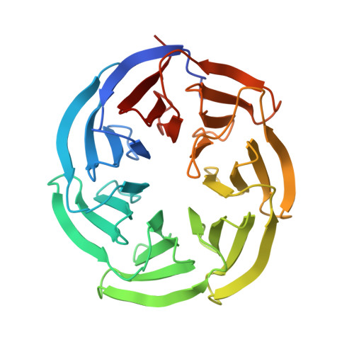
Structure of the WD40-domain of human ATG16L1.

Bajagic, M.Archna, A.Busing, P.Scrima, A.

(2017) Protein Sci 26: 1828-1837

  • DOI: https://doi.org/10.1002/pro.3222
  • Primary Citation of Related Structures:  
    5NUV

  • PubMed Abstract: 

    Autophagy-related protein ATG16L1 is a component of the mammalian ATG12∼ATG5/ATG16L1 complex, which acts as E3-ligase to catalyze lipidation of LC3 during autophagosome biogenesis. The N-terminal part of ATG16L1 comprises the ATG5-binding site and coiled-coil dimerization domain, both also present in yeast ATG16 and essential for bulk and starvation induced autophagy. While absent in yeast ATG16, mammalian ATG16L1 further contains a predicted C-terminal WD40-domain, which has been shown to be involved in mediating interaction with diverse factors in the context of alternative functions of autophagy, such as inflammatory control and xenophagy. In this work, we provide detailed information on the domain boundaries of the WD40-domain of human ATG16L1 and present its crystal structure at a resolution of 1.55 Å.


  • Organizational Affiliation

    Structural Biology of Autophagy Group, Department of Structure and Function of Proteins, Helmholtz Centre for Infection Research, Braunschweig, 38124, Germany.


Macromolecules
Find similar proteins by:  (by identity cutoff)  |  3D Structure
Entity ID: 1
MoleculeChains Sequence LengthOrganismDetailsImage
Autophagy-related protein 16-1309Homo sapiensMutation(s): 3 
Gene Names: ATG16L1APG16LUNQ9393/PRO34307
UniProt & NIH Common Fund Data Resources
Find proteins for Q676U5 (Homo sapiens)
Explore Q676U5 
Go to UniProtKB:  Q676U5
PHAROS:  Q676U5
GTEx:  ENSG00000085978 
Entity Groups  
Sequence Clusters30% Identity50% Identity70% Identity90% Identity95% Identity100% Identity
UniProt GroupQ676U5
Sequence Annotations
Expand
  • Reference Sequence
Small Molecules
Ligands 1 Unique
IDChains Name / Formula / InChI Key2D Diagram3D Interactions
BU3
Query on BU3

Download Ideal Coordinates CCD File 
B [auth A](R,R)-2,3-BUTANEDIOL
C4 H10 O2
OWBTYPJTUOEWEK-QWWZWVQMSA-N
Experimental Data & Validation

Experimental Data

  • Method: X-RAY DIFFRACTION
  • Resolution: 1.55 Å
  • R-Value Free: 0.197 
  • R-Value Work: 0.165 
  • R-Value Observed: 0.167 
  • Space Group: C 1 2 1
Unit Cell:
Length ( Å )Angle ( ˚ )
a = 157.55α = 90
b = 38.22β = 105.74
c = 45.62γ = 90
Software Package:
Software NamePurpose
XSCALEdata scaling
PHASERphasing
PHENIXrefinement
PDB_EXTRACTdata extraction
XDSdata reduction

Structure Validation

View Full Validation Report


Entry History & Funding Information

Deposition Data


Funding OrganizationLocationGrant Number
Helmholtz AssociationGermanyVH-NG-727

Revision History  (Full details and data files)

  • Version 1.0: 2017-07-19
    Type: Initial release
  • Version 1.1: 2017-09-06
    Changes: Database references
  • Version 1.2: 2017-10-11
    Changes: Data collection
  • Version 1.3: 2024-01-17
    Changes: Data collection, Database references, Refinement description