5NQG

Crystal structure of Plasmodium vivax AMA1 in complex with a 39 aa PvRON2 peptide


Experimental Data Snapshot

  • Method: X-RAY DIFFRACTION
  • Resolution: 2.15 Å
  • R-Value Free: 0.226 
  • R-Value Work: 0.174 
  • R-Value Observed: 0.177 

wwPDB Validation   3D Report Full Report


This is version 1.1 of the entry. See complete history


Literature

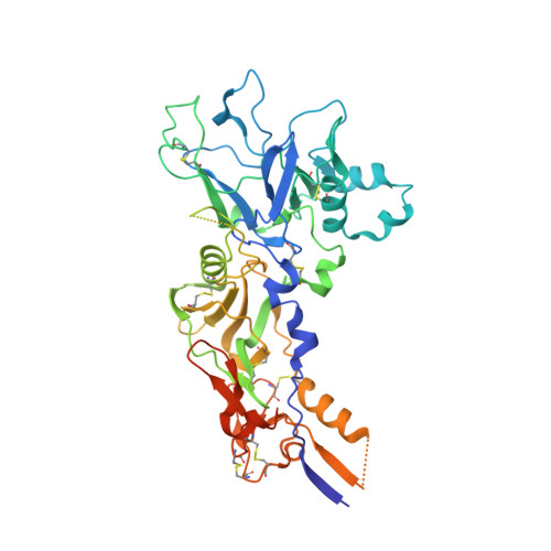
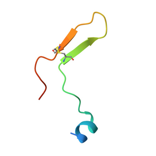
Cross-reactivity between apical membrane antgen 1 and rhoptry neck protein 2 in P. vivax and P. falciparum: A structural and binding study.

Vulliez-Le Normand, B.Saul, F.A.Hoos, S.Faber, B.W.Bentley, G.A.

(2017) PLoS One 12: e0183198-e0183198

  • DOI: https://doi.org/10.1371/journal.pone.0183198
  • Primary Citation of Related Structures:  
    5NQF, 5NQG

  • PubMed Abstract: 

    Malaria, a disease endemic in many tropical and subtropical regions, is caused by infection of the erythrocyte by the apicomplexan parasite Plasmodium. Host-cell invasion is a complex process but two Plasmodium proteins, Apical Membrane Antigen 1 (AMA1) and the Rhoptry Neck protein complex (RON), play a key role. AMA1, present on the surface of the parasite, binds tightly to the RON2 component of the RON protein complex, which is inserted into the erythrocyte membrane during invasion. Blocking the AMA1-RON2 interaction with antibodies or peptides inhibits invasion, underlining its importance in the Plasmodium life cycle and as a target for therapeutic strategies. We describe the crystal structure of the complex formed between AMA1 from P. vivax (PvAMA1) and a peptide derived from the externally exposed region of P. vivax RON2 (PvRON2sp1), and of the heterocomplex formed between P. falciparum AMA1 (PfAMA1) and PvRON2sp1. Binding studies show that the affinity of PvRON2sp1 for PvAMA1 is weaker than that previously reported for the PfRON2sp1-PfAMA1 association. Moreover, while PvRON2sp1 shows strong cross-reactivity with PfAMA1, PfRON2sp1 displays no detectable interaction with PvAMA1. The structures show that the equivalent residues PvRON2-Thr2055 and PfRON2-Arg2041 largely account for this pattern of reactivity.


  • Organizational Affiliation

    Institut Pasteur, Unité de Microbiologie Structurale, Département de Biologie Structurale et Chimie, Centre National de la Recherche Scientifique, UMR 3528, Université Paris Diderot, Sorbonne Paris Cité, Microbiologie Structurale, Paris, France.


Macromolecules
Find similar proteins by:  (by identity cutoff)  |  3D Structure
Entity ID: 1
MoleculeChains Sequence LengthOrganismDetailsImage
Apical merozoite antigen 1471Plasmodium vivax Sal-1Mutation(s): 4 
Gene Names: AMA1
UniProt
Find proteins for A5K4Z2 (Plasmodium vivax (strain Salvador I))
Explore A5K4Z2 
Go to UniProtKB:  A5K4Z2
Entity Groups  
Sequence Clusters30% Identity50% Identity70% Identity90% Identity95% Identity100% Identity
UniProt GroupA5K4Z2
Sequence Annotations
Expand
  • Reference Sequence
Find similar proteins by:  (by identity cutoff)  |  3D Structure
Entity ID: 2
MoleculeChains Sequence LengthOrganismDetailsImage
RON239Plasmodium vivaxMutation(s): 0 
UniProt
Find proteins for A5K3N8 (Plasmodium vivax (strain Salvador I))
Explore A5K3N8 
Go to UniProtKB:  A5K3N8
Entity Groups  
Sequence Clusters30% Identity50% Identity70% Identity90% Identity95% Identity100% Identity
UniProt GroupA5K3N8
Sequence Annotations
Expand
  • Reference Sequence
Experimental Data & Validation

Experimental Data

  • Method: X-RAY DIFFRACTION
  • Resolution: 2.15 Å
  • R-Value Free: 0.226 
  • R-Value Work: 0.174 
  • R-Value Observed: 0.177 
  • Space Group: C 1 2 1
Unit Cell:
Length ( Å )Angle ( ˚ )
a = 164.46α = 90
b = 54β = 105.71
c = 62.13γ = 90
Software Package:
Software NamePurpose
BUSTERrefinement
XDSdata reduction
SCALAdata scaling
PHASERphasing

Structure Validation

View Full Validation Report


Entry History 

Deposition Data

Revision History  (Full details and data files)

  • Version 1.0: 2017-09-06
    Type: Initial release
  • Version 1.1: 2024-01-17
    Changes: Data collection, Database references, Refinement description