5NP0

Closed dimer of human ATM (Ataxia telangiectasia mutated)


Experimental Data Snapshot

  • Method: ELECTRON MICROSCOPY
  • Resolution: 5.70 Å
  • Aggregation State: PARTICLE 
  • Reconstruction Method: SINGLE PARTICLE 

wwPDB Validation   3D Report Full Report


This is version 1.6 of the entry. See complete history


Literature

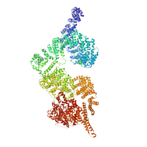
Structures of closed and open conformations of dimeric human ATM.

Baretic, D.Pollard, H.K.Fisher, D.I.Johnson, C.M.Santhanam, B.Truman, C.M.Kouba, T.Fersht, A.R.Phillips, C.Williams, R.L.

(2017) Sci Adv 3: e1700933-e1700933

  • DOI: https://doi.org/10.1126/sciadv.1700933
  • Primary Citation of Related Structures:  
    5NP0, 5NP1

  • PubMed Abstract: 

    ATM (ataxia-telangiectasia mutated) is a phosphatidylinositol 3-kinase-related protein kinase (PIKK) best known for its role in DNA damage response. ATM also functions in oxidative stress response, insulin signaling, and neurogenesis. Our electron cryomicroscopy (cryo-EM) suggests that human ATM is in a dynamic equilibrium between closed and open dimers. In the closed state, the PIKK regulatory domain blocks the peptide substrate-binding site, suggesting that this conformation may represent an inactive or basally active enzyme. The active site is held in this closed conformation by interaction with a long helical hairpin in the TRD3 (tetratricopeptide repeats domain 3) domain of the symmetry-related molecule. The open dimer has two protomers with only a limited contact interface, and it lacks the intermolecular interactions that block the peptide-binding site in the closed dimer. This suggests that the open conformation may be more active. The ATM structure shows the detailed topology of the regulator-interacting N-terminal helical solenoid. The ATM conformational dynamics shown by the structures represent an important step in understanding the enzyme regulation.


  • Organizational Affiliation

    Medical Research Council Laboratory of Molecular Biology, Cambridge CB2 0QH, UK.


Macromolecules
Find similar proteins by:  (by identity cutoff)  |  3D Structure
Entity ID: 1
MoleculeChains Sequence LengthOrganismDetailsImage
Serine-protein kinase ATM
A, B
3,066Homo sapiensMutation(s): 0 
Gene Names: ATM
EC: 2.7.11.1
UniProt & NIH Common Fund Data Resources
Find proteins for Q13315 (Homo sapiens)
Explore Q13315 
Go to UniProtKB:  Q13315
PHAROS:  Q13315
GTEx:  ENSG00000149311 
Entity Groups  
Sequence Clusters30% Identity50% Identity70% Identity90% Identity95% Identity100% Identity
UniProt GroupQ13315
Sequence Annotations
Expand
  • Reference Sequence
Experimental Data & Validation

Experimental Data

  • Method: ELECTRON MICROSCOPY
  • Resolution: 5.70 Å
  • Aggregation State: PARTICLE 
  • Reconstruction Method: SINGLE PARTICLE 
EM Software:
TaskSoftware PackageVersion
MODEL REFINEMENTPHENIX1.10.1_2155
RECONSTRUCTIONRELION1.4

Structure Validation

View Full Validation Report


Entry History & Funding Information

Deposition Data


Funding OrganizationLocationGrant Number
Medical Research Council (United Kingdom)United KingdomMC_U105184308
LMB/AstraZenecaUnited KingdomMC_A024-5PF9H

Revision History  (Full details and data files)

  • Version 1.0: 2017-05-17
    Type: Initial release
  • Version 1.1: 2017-05-31
    Changes: Database references
  • Version 1.2: 2017-07-26
    Changes: Data collection, Experimental preparation
  • Version 1.3: 2017-08-30
    Changes: Author supporting evidence, Data collection
  • Version 1.4: 2018-01-31
    Changes: Data processing, Experimental preparation, Other
  • Version 1.5: 2018-02-07
    Changes: Data collection
  • Version 1.6: 2019-12-11
    Changes: Other