5NOC

Solution NMR Structure of the C-terminal domain of ParB (Spo0J)


Experimental Data Snapshot

  • Method: SOLUTION NMR
  • Conformers Calculated: 200 
  • Conformers Submitted: 20 
  • Selection Criteria: structures with the lowest energy 

wwPDB Validation   3D Report Full Report


This is version 1.3 of the entry. See complete history


Literature

The structural basis for dynamic DNA binding and bridging interactions which condense the bacterial centromere.

Fisher, G.L.Pastrana, C.L.Higman, V.A.Koh, A.Taylor, J.A.Butterer, A.Craggs, T.Sobott, F.Murray, H.Crump, M.P.Moreno-Herrero, F.Dillingham, M.S.

(2017) Elife 6

  • DOI: https://doi.org/10.7554/eLife.28086
  • Primary Citation of Related Structures:  
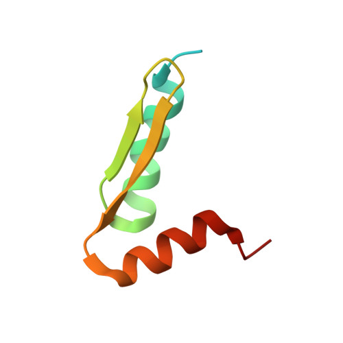
    5NOC

  • PubMed Abstract: 

    The ParB protein forms DNA bridging interactions around parS to condense DNA and earmark the bacterial chromosome for segregation. The molecular mechanism underlying the formation of these ParB networks is unclear. We show here that while the central DNA binding domain is essential for anchoring at parS , this interaction is not required for DNA condensation. Structural analysis of the C-terminal domain reveals a dimer with a lysine-rich surface that binds DNA non-specifically and is essential for DNA condensation in vitro. Mutation of either the dimerisation or the DNA binding interface eliminates ParB-GFP foci formation in vivo. Moreover, the free C-terminal domain can rapidly decondense ParB networks independently of its ability to bind DNA. Our work reveals a dual role for the C-terminal domain of ParB as both a DNA binding and bridging interface, and highlights the dynamic nature of ParB networks in Bacillus subtilis .


  • Organizational Affiliation

    DNA:protein Interactions Unit, School of Biochemistry, University of Bristol, Bristol, United Kingdom.


Macromolecules
Find similar proteins by:  (by identity cutoff)  |  3D Structure
Entity ID: 1
MoleculeChains Sequence LengthOrganismDetailsImage
Stage 0 sporulation protein J
A, B
69Bacillus subtilis subsp. subtilis str. 168Mutation(s): 0 
Gene Names: spo0JBSU40960
UniProt
Find proteins for P26497 (Bacillus subtilis (strain 168))
Explore P26497 
Go to UniProtKB:  P26497
Entity Groups  
Sequence Clusters30% Identity50% Identity70% Identity90% Identity95% Identity100% Identity
UniProt GroupP26497
Sequence Annotations
Expand
  • Reference Sequence
Experimental Data & Validation

Experimental Data

  • Method: SOLUTION NMR
  • Conformers Calculated: 200 
  • Conformers Submitted: 20 
  • Selection Criteria: structures with the lowest energy 

Structure Validation

View Full Validation Report


Entry History & Funding Information

Deposition Data


Funding OrganizationLocationGrant Number
Wellcome TrustUnited Kingdom100401/Z/12/Z
Biotechnology and Biological Sciences Research CouncilUnited Kingdom1363883

Revision History  (Full details and data files)

  • Version 1.0: 2017-12-13
    Type: Initial release
  • Version 1.1: 2017-12-27
    Changes: Database references
  • Version 1.2: 2019-05-08
    Changes: Data collection
  • Version 1.3: 2023-06-14
    Changes: Database references, Other