5NL7

The crystal structure of the Actin Binding Domain (ABD) of alpha actinin from Entamoeba histolytica


Experimental Data Snapshot

  • Method: X-RAY DIFFRACTION
  • Resolution: 2.48 Å
  • R-Value Free: 0.252 
  • R-Value Work: 0.196 
  • R-Value Observed: 0.199 

wwPDB Validation   3D Report Full Report


This is version 1.2 of the entry. See complete history


Literature

Calcium modulates the domain flexibility and function of an alpha-actinin similar to the ancestral alpha-actinin.

Pinotsis, N.Zielinska, K.Babuta, M.Arolas, J.L.Kostan, J.Khan, M.B.Schreiner, C.Salmazo, A.Ciccarelli, L.Puchinger, M.Gkougkoulia, E.A.Ribeiro Jr., E.A.Marlovits, T.C.Bhattacharya, A.Djinovic-Carugo, K.

(2020) Proc Natl Acad Sci U S A 117: 22101-22112

  • DOI: https://doi.org/10.1073/pnas.1917269117
  • Primary Citation of Related Structures:  
    5NL6, 5NL7, 6SL2, 6SL3, 6SL7

  • PubMed Abstract: 

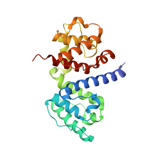
    The actin cytoskeleton, a dynamic network of actin filaments and associated F-actin-binding proteins, is fundamentally important in eukaryotes. α-Actinins are major F-actin bundlers that are inhibited by Ca 2+ in nonmuscle cells. Here we report the mechanism of Ca 2+ -mediated regulation of Entamoeba histolytica α-actinin-2 ( Eh Actn2) with features expected for the common ancestor of Entamoeba and higher eukaryotic α-actinins. Crystal structures of Ca 2+ -free and Ca 2+ -bound Eh Actn2 reveal a calmodulin-like domain (CaMD) uniquely inserted within the rod domain. Integrative studies reveal an exceptionally high affinity of the Eh Actn2 CaMD for Ca 2+ , binding of which can only be regulated in the presence of physiological concentrations of Mg 2+ Ca 2+ binding triggers an increase in protein multidomain rigidity, reducing conformational flexibility of F-actin-binding domains via interdomain cross-talk and consequently inhibiting F-actin bundling. In vivo studies uncover that Eh Actn2 plays an important role in phagocytic cup formation and might constitute a new drug target for amoebic dysentery.


  • Organizational Affiliation

    Department of Structural and Computational Biology, Max Perutz Labs, University of Vienna, A-1030 Vienna, Austria.


Macromolecules
Find similar proteins by:  (by identity cutoff)  |  3D Structure
Entity ID: 1
MoleculeChains Sequence LengthOrganismDetailsImage
Calponin homology domain protein putative
A, B
234Entamoeba histolyticaMutation(s): 0 
Gene Names: CL6EHI_199000EHI_199000
UniProt
Find proteins for C4LWU6 (Entamoeba histolytica (strain ATCC 30459 / HM-1:IMSS / ABRM))
Explore C4LWU6 
Go to UniProtKB:  C4LWU6
Entity Groups  
Sequence Clusters30% Identity50% Identity70% Identity90% Identity95% Identity100% Identity
UniProt GroupC4LWU6
Sequence Annotations
Expand
  • Reference Sequence
Small Molecules
Ligands 2 Unique
IDChains Name / Formula / InChI Key2D Diagram3D Interactions
TRS
Query on TRS

Download Ideal Coordinates CCD File 
D [auth A]2-AMINO-2-HYDROXYMETHYL-PROPANE-1,3-DIOL
C4 H12 N O3
LENZDBCJOHFCAS-UHFFFAOYSA-O
CA
Query on CA

Download Ideal Coordinates CCD File 
C [auth A]CALCIUM ION
Ca
BHPQYMZQTOCNFJ-UHFFFAOYSA-N
Experimental Data & Validation

Experimental Data

  • Method: X-RAY DIFFRACTION
  • Resolution: 2.48 Å
  • R-Value Free: 0.252 
  • R-Value Work: 0.196 
  • R-Value Observed: 0.199 
  • Space Group: P 3
Unit Cell:
Length ( Å )Angle ( ˚ )
a = 146.74α = 90
b = 146.74β = 90
c = 33.97γ = 120
Software Package:
Software NamePurpose
PHENIXrefinement
XDSdata reduction
XSCALEdata scaling
MOLREPphasing

Structure Validation

View Full Validation Report


Entry History 

Deposition Data

Revision History  (Full details and data files)

  • Version 1.0: 2018-05-16
    Type: Initial release
  • Version 1.1: 2020-11-04
    Changes: Database references, Derived calculations
  • Version 1.2: 2024-01-17
    Changes: Data collection, Database references, Refinement description