5NIM

EthR complex


Experimental Data Snapshot

  • Method: X-RAY DIFFRACTION
  • Resolution: 1.50 Å
  • R-Value Free: 0.204 
  • R-Value Work: 0.175 
  • R-Value Observed: 0.177 

wwPDB Validation   3D Report Full Report


Ligand Structure Quality Assessment 


This is version 1.2 of the entry. See complete history


Literature

New active leads for tuberculosis booster drugs by structure-based drug discovery.

Tatum, N.J.Liebeschuetz, J.W.Cole, J.C.Frita, R.Herledan, A.Baulard, A.R.Willand, N.Pohl, E.

(2017) Org Biomol Chem 15: 10245-10255

  • DOI: https://doi.org/10.1039/c7ob00910k
  • Primary Citation of Related Structures:  
    5NIM, 5NIO, 5NIZ, 5NJ0

  • PubMed Abstract: 

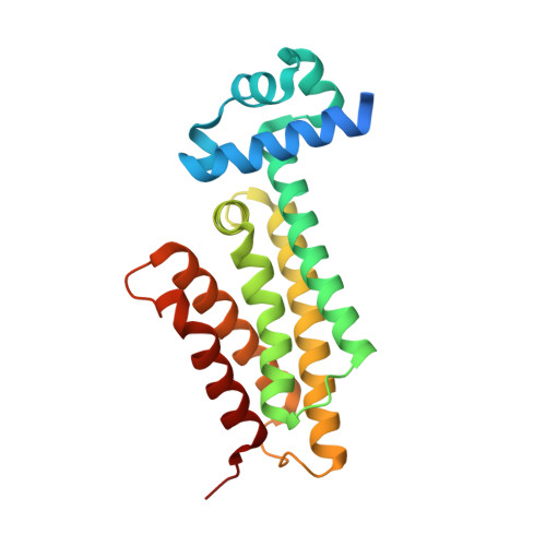
    The transcriptional repressor EthR from Mycobacterium tuberculosis, a member of the TetR family of prokaryotic homo-dimeric transcription factors, controls the expression of the mycobacterial mono-oxygenase EthA. EthA is responsible for the bio-activation of the second-line tuberculosis pro-drug ethionamide, and consequently EthR inhibitors boost drug efficacy. Here, we present a comprehensive in silico structure-based screening protocol that led to the identification of a number of novel scaffolds of EthR inhibitors in subsequent biophysical screening by thermal shift assay. Growth inhibition assays demonstrated that five of the twenty biophysical hits were capable of boosting ethionamide activity in vitro, with the best novel scaffold displaying an EC 50 of 34 μM. In addition, the co-crystal structures of EthR with four new ligands at resolution ranging from 2.1 to 1.4 Å confirm the binding and inactivation mode, and will enable future lead development.


  • Organizational Affiliation

    Department of Chemistry, Durham University, South Road, Durham DH1 3LE, UK. ehmke.pohl@durham.ac.uk.


Macromolecules
Find similar proteins by:  (by identity cutoff)  |  3D Structure
Entity ID: 1
MoleculeChains Sequence LengthOrganismDetailsImage
HTH-type transcriptional regulator EthR216Mycobacterium tuberculosisMutation(s): 0 
Gene Names: ethRetaRRv3855
UniProt
Find proteins for P9WMC1 (Mycobacterium tuberculosis (strain ATCC 25618 / H37Rv))
Explore P9WMC1 
Go to UniProtKB:  P9WMC1
Entity Groups  
Sequence Clusters30% Identity50% Identity70% Identity90% Identity95% Identity100% Identity
UniProt GroupP9WMC1
Sequence Annotations
Expand
  • Reference Sequence
Small Molecules
Ligands 2 Unique
IDChains Name / Formula / InChI Key2D Diagram3D Interactions
8YE
Query on 8YE

Download Ideal Coordinates CCD File 
B [auth A][1-(2-hydroxyethyl)pyrrolo[3,4-c]pyrazol-5-yl]-(5-propyl-1,2-oxazol-3-yl)methanone
C14 H16 N4 O3
NYKTWGKCNASWSV-UHFFFAOYSA-N
SO4
Query on SO4

Download Ideal Coordinates CCD File 
C [auth A]SULFATE ION
O4 S
QAOWNCQODCNURD-UHFFFAOYSA-L
Experimental Data & Validation

Experimental Data

  • Method: X-RAY DIFFRACTION
  • Resolution: 1.50 Å
  • R-Value Free: 0.204 
  • R-Value Work: 0.175 
  • R-Value Observed: 0.177 
  • Space Group: P 41 21 2
Unit Cell:
Length ( Å )Angle ( ˚ )
a = 122.544α = 90
b = 122.544β = 90
c = 33.815γ = 90
Software Package:
Software NamePurpose
REFMACrefinement
xia2data reduction
xia2data scaling
PHASERphasing

Structure Validation

View Full Validation Report


Ligand Structure Quality Assessment 


Entry History 

Deposition Data

Revision History  (Full details and data files)

  • Version 1.0: 2017-11-15
    Type: Initial release
  • Version 1.1: 2017-12-06
    Changes: Database references
  • Version 1.2: 2017-12-27
    Changes: Database references