5NG9

Crystal structure of the GluA2 ligand-binding domain (S1S2J) in complex with agonist CIP-AS at 1.15 A resolution.


Experimental Data Snapshot

  • Method: X-RAY DIFFRACTION
  • Resolution: 1.15 Å
  • R-Value Free: 0.133 
  • R-Value Work: 0.109 
  • R-Value Observed: 0.110 

wwPDB Validation   3D Report Full Report


Ligand Structure Quality Assessment 


This is version 1.2 of the entry. See complete history


Literature

Structure and Affinity of Two Bicyclic Glutamate Analogues at AMPA and Kainate Receptors.

Mllerud, S.Pinto, A.Marconi, L.Frydenvang, K.Thorsen, T.S.Laulumaa, S.Venskutonyte, R.Winther, S.Moral, A.M.C.Tamborini, L.Conti, P.Pickering, D.S.Kastrup, J.S.

(2017) ACS Chem Neurosci 8: 2056-2064

  • DOI: https://doi.org/10.1021/acschemneuro.7b00201
  • Primary Citation of Related Structures:  
    5NEB, 5NF5, 5NF6, 5NG9, 5NIH, 5O4F

  • PubMed Abstract: 

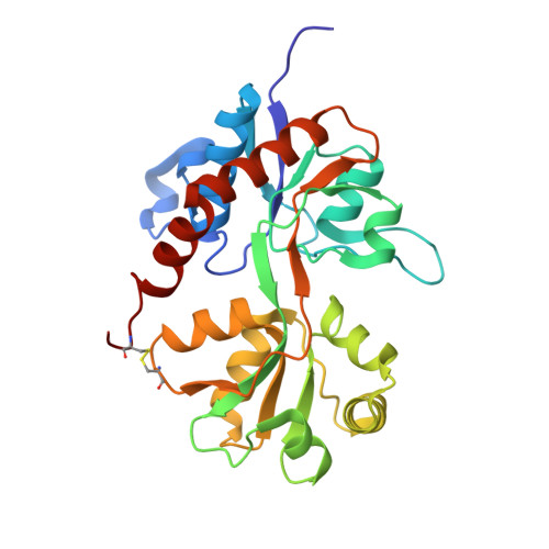
    Ionotropic glutamate receptors (iGluRs) are involved in most of the fast excitatory synaptic transmission in the central nervous system. These receptors are important for learning and memory formation, but are also involved in the development of diseases such as Alzheimer's disease, epilepsy and depression. To understand the function of different types of iGluRs, selective agonists are invaluable as pharmacological tool compounds. Here, we report binding affinities of two bicyclic, conformationally restricted analogues of glutamate (CIP-AS and LM-12b) at AMPA (GluA2 and GluA3) and kainate receptor subunits (GluK1-3 and GluK5). Both CIP-AS and LM-12b were found to be GluK3-preferring agonists, with K i of 6 and 22 nM, respectively, at recombinant GluK3 receptors. The detailed binding mode of CIP-AS and LM-12b in the ligand-binding domains of the AMPA receptor subunit GluA2 (GluA2-LBD) and the kainate receptor subunits GluK1 (GluK1-LBD) and GluK3 (GluK3-LBD) was investigated by X-ray crystallography. CIP-AS stabilized all three receptor constructs in conformations similar to those with kainate. Remarkably, whereas LM-12b bound in a similar manner to CIP-AS in GluA2-LBD and GluK3-LBD, it introduced full closure of the ligand-binding domain in GluK1-LBD and formation of a D1-D2 interlobe hydrogen bond between Glu441 and Ser721, as also observed with glutamate. As the binding affinity of LM-12b at GluK1 is ∼8-fold better than that for CIP-AS (K i of 85 and 656 nM, respectively), it shows that small changes in agonist structure can lead to prominent differences in structure and function.


  • Organizational Affiliation

    Department of Drug Design and Pharmacology, Faculty of Health and Medical Sciences, University of Copenhagen , 2100 Copenhagen, Denmark.


Macromolecules
Find similar proteins by:  (by identity cutoff)  |  3D Structure
Entity ID: 1
MoleculeChains Sequence LengthOrganismDetailsImage
Glutamate receptor 2264Rattus norvegicusMutation(s): 0 
Gene Names: Gria2Glur2
UniProt
Find proteins for P19491 (Rattus norvegicus)
Explore P19491 
Go to UniProtKB:  P19491
Entity Groups  
Sequence Clusters30% Identity50% Identity70% Identity90% Identity95% Identity100% Identity
UniProt GroupP19491
Sequence Annotations
Expand
  • Reference Sequence
Small Molecules
Ligands 6 Unique
IDChains Name / Formula / InChI Key2D Diagram3D Interactions
8WQ
Query on 8WQ

Download Ideal Coordinates CCD File 
F [auth A](2~{S},3~{R},4~{R})-3-(carboxycarbonyl)-4-oxidanyl-pyrrolidine-2-carboxylic acid
C7 H9 N O6
XAWWSRPWEJXFEU-HZLVTQRSSA-N
8VN
Query on 8VN

Download Ideal Coordinates CCD File 
E [auth A](3~{a}~{S},4~{S},6~{a}~{R})-4,5,6,6~{a}-tetrahydro-3~{a}~{H}-pyrrolo[3,4-d][1,2]oxazole-3,4-dicarboxylic acid
C7 H8 N2 O5
JMTZNIDIUDLCMK-HZLVTQRSSA-N
FLC
Query on FLC

Download Ideal Coordinates CCD File 
B [auth A],
C [auth A]
CITRATE ANION
C6 H5 O7
KRKNYBCHXYNGOX-UHFFFAOYSA-K
SO4
Query on SO4

Download Ideal Coordinates CCD File 
G [auth A],
H [auth A],
I [auth A]
SULFATE ION
O4 S
QAOWNCQODCNURD-UHFFFAOYSA-L
GOL
Query on GOL

Download Ideal Coordinates CCD File 
J [auth A]GLYCEROL
C3 H8 O3
PEDCQBHIVMGVHV-UHFFFAOYSA-N
LI
Query on LI

Download Ideal Coordinates CCD File 
D [auth A]LITHIUM ION
Li
HBBGRARXTFLTSG-UHFFFAOYSA-N
Binding Affinity Annotations 
IDSourceBinding Affinity
8VN Binding MOAD:  5NG9 Ki: 191 (nM) from 1 assay(s)
Experimental Data & Validation

Experimental Data

  • Method: X-RAY DIFFRACTION
  • Resolution: 1.15 Å
  • R-Value Free: 0.133 
  • R-Value Work: 0.109 
  • R-Value Observed: 0.110 
  • Space Group: P 21 21 2
Unit Cell:
Length ( Å )Angle ( ˚ )
a = 62.218α = 90
b = 88.14β = 90
c = 47.958γ = 90
Software Package:
Software NamePurpose
PHENIXrefinement
SCALAdata scaling
PDB_EXTRACTdata extraction
XDSdata reduction
PHASERphasing

Structure Validation

View Full Validation Report


Ligand Structure Quality Assessment 


Entry History 

Deposition Data

Revision History  (Full details and data files)

  • Version 1.0: 2017-07-26
    Type: Initial release
  • Version 1.1: 2017-09-27
    Changes: Database references
  • Version 1.2: 2024-01-17
    Changes: Data collection, Database references, Refinement description