5NC0

The 0.91 A resolution structure of the L16G mutant of cytochrome c prime from Alcaligenes xylosoxidans, complexed with nitric oxide


Experimental Data Snapshot

  • Method: X-RAY DIFFRACTION
  • Resolution: 0.91 Å
  • R-Value Free: 0.132 
  • R-Value Work: 0.125 
  • R-Value Observed: 0.125 

wwPDB Validation   3D Report Full Report


Ligand Structure Quality Assessment 


This is version 2.1 of the entry. See complete history


Literature

Distinguishing Nitro vs Nitrito Coordination in Cytochrome c' Using Vibrational Spectroscopy and Density Functional Theory.

Nilsson, Z.N.Mandella, B.L.Sen, K.Kekilli, D.Hough, M.A.Moenne-Loccoz, P.Strange, R.W.Andrew, C.R.

(2017) Inorg Chem 56: 13205-13213

  • DOI: https://doi.org/10.1021/acs.inorgchem.7b01945
  • Primary Citation of Related Structures:  
    5NC0, 5NGX

  • PubMed Abstract: 

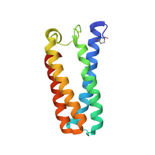
    Nitrite coordination to heme cofactors is a key step in the anaerobic production of the signaling molecule nitric oxide (NO). An ambidentate ligand, nitrite has the potential to coordinate via the N- (nitro) or O- (nitrito) atoms in a manner that can direct its reactivity. Distinguishing nitro vs nitrito coordination, along with the influence of the surrounding protein, is therefore of particular interest. In this study, we probed Fe(III) heme-nitrite coordination in Alcaligenes xylosoxidans cytochrome c' (AXCP), an NO carrier that excludes anions in its native state but that readily binds nitrite (K d ∼ 0.5 mM) following a distal Leu16 → Gly mutation to remove distal steric constraints. Room-temperature resonance Raman spectra (407 nm excitation) identify ν(Fe-NO 2 ), δ(ONO), and ν s (NO 2 ) nitrite ligand vibrations in solution. Illumination with 351 nm UV light results in photoconversion to {FeNO} 6 and {FeNO} 7 states, enabling FTIR measurements to distinguish ν s (NO 2 ) and ν as (NO 2 ) vibrations from differential spectra. Density functional theory calculations highlight the connections between heme environment, nitrite coordination mode, and vibrational properties and confirm that nitrite binds to L16G AXCP exclusively through the N atom. Efforts to obtain the nitrite complex crystal structure were hampered by photochemistry in the X-ray beam. Although low dose crystal structures could be modeled with a mixed nitrite (nitro)/H 2 O distal population, their photosensitivity and partial occupancy underscores the value of the vibrational approach. Overall, this study sheds light on steric determinants of heme-nitrite binding and provides vibrational benchmarks for future studies of heme protein nitrite reactions.


  • Organizational Affiliation

    Department of Chemistry and Biochemistry, Eastern Oregon University , La Grande, Oregon 97850, United States.


Macromolecules
Find similar proteins by:  (by identity cutoff)  |  3D Structure
Entity ID: 1
MoleculeChains Sequence LengthOrganismDetailsImage
Cytochrome c'127Achromobacter xylosoxidansMutation(s): 1 
UniProt
Find proteins for P00138 (Alcaligenes xylosoxydans xylosoxydans)
Explore P00138 
Go to UniProtKB:  P00138
Entity Groups  
Sequence Clusters30% Identity50% Identity70% Identity90% Identity95% Identity100% Identity
UniProt GroupP00138
Sequence Annotations
Expand
  • Reference Sequence
Small Molecules
Ligands 6 Unique
IDChains Name / Formula / InChI Key2D Diagram3D Interactions
HEC
Query on HEC

Download Ideal Coordinates CCD File 
B [auth A]HEME C
C34 H34 Fe N4 O4
HXQIYSLZKNYNMH-LJNAALQVSA-N
SO4
Query on SO4

Download Ideal Coordinates CCD File 
F [auth A],
G [auth A],
H [auth A],
I [auth A]
SULFATE ION
O4 S
QAOWNCQODCNURD-UHFFFAOYSA-L
EDO
Query on EDO

Download Ideal Coordinates CCD File 
K [auth A],
L [auth A],
M [auth A]
1,2-ETHANEDIOL
C2 H6 O2
LYCAIKOWRPUZTN-UHFFFAOYSA-N
NO3
Query on NO3

Download Ideal Coordinates CCD File 
D [auth A]NITRATE ION
N O3
NHNBFGGVMKEFGY-UHFFFAOYSA-N
NO2
Query on NO2

Download Ideal Coordinates CCD File 
E [auth A],
J [auth A]
NITRITE ION
N O2
IOVCWXUNBOPUCH-UHFFFAOYSA-M
NO
Query on NO

Download Ideal Coordinates CCD File 
C [auth A]NITRIC OXIDE
N O
ODUCDPQEXGNKDN-UHFFFAOYSA-N
Modified Residues  1 Unique
IDChains TypeFormula2D DiagramParent
PCA
Query on PCA
A
L-PEPTIDE LINKINGC5 H7 N O3GLN
Experimental Data & Validation

Experimental Data

  • Method: X-RAY DIFFRACTION
  • Resolution: 0.91 Å
  • R-Value Free: 0.132 
  • R-Value Work: 0.125 
  • R-Value Observed: 0.125 
  • Space Group: P 65 2 2
Unit Cell:
Length ( Å )Angle ( ˚ )
a = 52.43α = 90
b = 52.43β = 90
c = 183.25γ = 120
Software Package:
Software NamePurpose
REFMACrefinement
XDSdata reduction
Aimlessdata scaling
REFMACphasing

Structure Validation

View Full Validation Report


Ligand Structure Quality Assessment 


Entry History 

Deposition Data

Revision History  (Full details and data files)

  • Version 1.0: 2018-01-10
    Type: Initial release
  • Version 2.0: 2020-03-11
    Changes: Polymer sequence
  • Version 2.1: 2024-01-17
    Changes: Data collection, Database references, Refinement description