5NBW

Crystal structure of the Fab fragment 22F12 in complex with 3-hydroxybenzo[a]pyrene


Experimental Data Snapshot

  • Method: X-RAY DIFFRACTION
  • Resolution: 2.40 Å
  • R-Value Free: 0.228 
  • R-Value Work: 0.180 
  • R-Value Observed: 0.183 

wwPDB Validation   3D Report Full Report


Ligand Structure Quality Assessment 


This is version 1.2 of the entry. See complete history


Literature

Tight Molecular Recognition of Benzo[a]pyrene by a High-Affinity Antibody.

Eichinger, A.Neumaier, I.Pschenitza, M.Niessner, R.Knopp, D.Skerra, A.

(2017) Angew Chem Int Ed Engl 56: 10592-10597

  • DOI: https://doi.org/10.1002/anie.201703893
  • Primary Citation of Related Structures:  
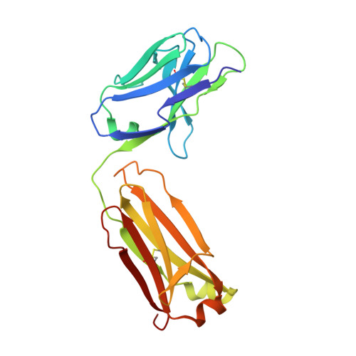
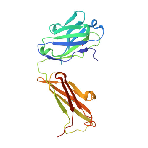
    5NBW

  • PubMed Abstract: 

    Benzo[a]pyrene, which is produced during the incomplete combustion of organic material, is an abundant noxious pollutant because of its carcinogenic metabolic degradation products. The high-affinity (K D ≈3 nm) monoclonal antibody 22F12 allows facile bioanalytical quantification of benzo[a]pyrene even in complex matrices. We report the functional and X-ray crystallographic analysis of 22F12 in complex with 3-hydroxybenzo[a]pyrene after cloning of the V-genes and production as a recombinant Fab fragment. The polycyclic aromatic hydrocarbon is bound in a deep pocket between the light and heavy chains, surrounded mainly by aromatic and aliphatic amino acid side chains. Interestingly, the hapten-antibody interface is less densely packed than expected and reveals polar, H-bond-like interactions with the polycyclic aromatic π-electron system, which may allow the antibody to maintain a large, predominantly hydrophobic binding site in an aqueous environment while providing sufficient complementarity to its ligand.


  • Organizational Affiliation

    Munich Center for Integrated Protein Science, CIPS-M, und Lehrstuhl für Biologische Chemie, Technische Universität München, 85354, Freising, Weihenstephan, Germany.


Macromolecules
Find similar proteins by:  (by identity cutoff)  |  3D Structure
Entity ID: 1
MoleculeChains Sequence LengthOrganismDetailsImage
Fab 22F12 (L,H)A [auth L],
C [auth A]
214Mus musculusMutation(s): 0 
Entity Groups  
Sequence Clusters30% Identity50% Identity70% Identity90% Identity95% Identity100% Identity
Sequence Annotations
Expand
  • Reference Sequence
Find similar proteins by:  (by identity cutoff)  |  3D Structure
Entity ID: 2
MoleculeChains Sequence LengthOrganismDetailsImage
Fab 22F12 (A,B)B [auth H],
D [auth B]
222Mus musculusMutation(s): 0 
Entity Groups  
Sequence Clusters30% Identity50% Identity70% Identity90% Identity95% Identity100% Identity
Sequence Annotations
Expand
  • Reference Sequence
Small Molecules
Experimental Data & Validation

Experimental Data

  • Method: X-RAY DIFFRACTION
  • Resolution: 2.40 Å
  • R-Value Free: 0.228 
  • R-Value Work: 0.180 
  • R-Value Observed: 0.183 
  • Space Group: P 21 21 21
Unit Cell:
Length ( Å )Angle ( ˚ )
a = 58.15α = 90
b = 81β = 90
c = 186.16γ = 90
Software Package:
Software NamePurpose
REFMACrefinement
MOSFLMdata reduction
SCALAdata scaling
PHASERphasing

Structure Validation

View Full Validation Report


Ligand Structure Quality Assessment 


Entry History 

Deposition Data

Revision History  (Full details and data files)

  • Version 1.0: 2017-06-28
    Type: Initial release
  • Version 1.1: 2017-08-30
    Changes: Database references
  • Version 1.2: 2024-01-17
    Changes: Data collection, Database references, Refinement description