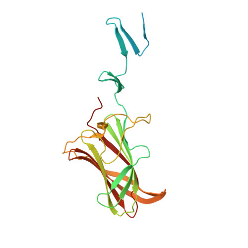
Structure and N-acetylglucosamine binding of the distal domain of mouse adenovirus 2 fibre.
Singh, A.K., Nguyen, T.H., Vidovszky, M.Z., Harrach, B., Benko, M., Kirwan, A., Joshi, L., Kilcoyne, M., Berbis, M.A., Canada, F.J., Jimenez-Barbero, J., Menendez, M., Wilson, S.S., Bromme, B.A., Smith, J.G., van Raaij, M.J.(2018) J Gen Virol 99: 1494-1508
- PubMed: 30277856
- DOI: https://doi.org/10.1099/jgv.0.001145
- Primary Citation of Related Structures:
5N83, 5N8D, 5NBH, 5NC1 - PubMed Abstract:
Murine adenovirus 2 (MAdV-2) infects cells of the mouse gastrointestinal tract. Like human adenoviruses, it is a member of the genus Mastadenovirus, family Adenoviridae. The MAdV-2 genome has a single fibre gene that expresses a 787 residue-long protein. Through analogy to other adenovirus fibre proteins, it is expected that the carboxy-terminal virus-distal head domain of the fibre is responsible for binding to the host cell, although the natural receptor is unknown. The putative head domain has little sequence identity to adenovirus fibres of known structure. In this report, we present high-resolution crystal structures of the carboxy-terminal part of the MAdV-2 fibre. The structures reveal a domain with the typical adenovirus fibre head topology and a domain containing two triple β-spiral repeats of the shaft domain. Through glycan microarray profiling, saturation transfer difference nuclear magnetic resonance spectroscopy, isothermal titration calorimetry and site-directed mutagenesis, we show that the fibre specifically binds to the monosaccharide N-acetylglucosamine (GlcNAc). The crystal structure of the complex reveals that GlcNAc binds between the AB and CD loops at the top of each of the three monomers of the MAdV-2 fibre head. However, infection competition assays show that soluble GlcNAc monosaccharide and natural GlcNAc-containing polymers do not inhibit infection by MAdV-2. Furthermore, site-directed mutation of the GlcNAc-binding residues does not prevent the inhibition of infection by soluble fibre protein. On the other hand, we show that the MAdV-2 fibre protein binds GlcNAc-containing mucin glycans, which suggests that the MAdV-2 fibre protein may play a role in viral mucin penetration in the mouse gut.
Organizational Affiliation:
1Departamento de Estructura de Macromoléculas, Centro Nacional de Biotecnologia (CNB-CSIC), Calle Darwin 3, 28049 Madrid, Spain.