5N9X

Structure of adenylation domain THR1 involved in the biosynthesis of 4-chlorothreonine in Streptomyces SP.OH-5093, ligand bound structure


Experimental Data Snapshot

  • Method: X-RAY DIFFRACTION
  • Resolution: 2.40 Å
  • R-Value Free: 0.276 
  • R-Value Work: 0.199 
  • R-Value Observed: 0.203 

wwPDB Validation   3D Report Full Report


Ligand Structure Quality Assessment 


This is version 1.2 of the entry. See complete history


Literature

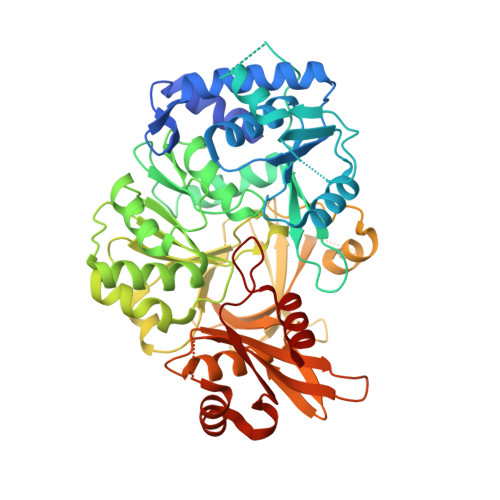
Structure of the adenylation domain Thr1 involved in the biosynthesis of 4-chlorothreonine in Streptomyces sp. OH-5093-protein flexibility and molecular bases of substrate specificity.

Scaglione, A.Fullone, M.R.Montemiglio, L.C.Parisi, G.Zamparelli, C.Vallone, B.Savino, C.Grgurina, I.

(2017) FEBS J 284: 2981-2999

  • DOI: https://doi.org/10.1111/febs.14163
  • Primary Citation of Related Structures:  
    5N9W, 5N9X

  • PubMed Abstract: 

    We determined the crystal structure of Thr1, the self-standing adenylation domain involved in the nonribosomal-like biosynthesis of free 4-chlorothreonine in Streptomyces sp. OH-5093. Thr1 shows two monomers in the crystallographic asymmetric unit with different relative orientations of the C- and N-terminal subdomains both in the presence of substrates and in the unliganded form. Cocrystallization with substrates, adenosine 5'-triphosphate and l-threonine, yielded one monomer containing the two substrates and the other in complex with l-threonine adenylate, locked in a postadenylation state. Steady-state kinetics showed that Thr1 activates l-Thr and its stereoisomers, as well as d-Ala, l- and d-Ser, albeit with lower efficiency. Modeling of these substrates in the active site highlighted the molecular bases of substrate discrimination. This work provides the first crystal structure of a threonine-activating adenylation enzyme, a contribution to the studies on conformational rearrangement in adenylation domains and on substrate recognition in nonribosomal biosynthesis.


  • Organizational Affiliation

    Department of Biochemical Sciences "A. Rossi Fanelli", Istituto Pasteur-Fondazione Cenci Bolognetti, Sapienza University of Rome, Italy.


Macromolecules
Find similar proteins by:  (by identity cutoff)  |  3D Structure
Entity ID: 1
MoleculeChains Sequence LengthOrganismDetailsImage
Adenylation domain
A, B
529Streptomyces sp.Mutation(s): 0 
Gene Names: thr1
UniProt
Find proteins for H6SG27 (Streptomyces sp)
Explore H6SG27 
Go to UniProtKB:  H6SG27
Entity Groups  
Sequence Clusters30% Identity50% Identity70% Identity90% Identity95% Identity100% Identity
UniProt GroupH6SG27
Sequence Annotations
Expand
  • Reference Sequence
Small Molecules
Ligands 4 Unique
IDChains Name / Formula / InChI Key2D Diagram3D Interactions
ATP
Query on ATP

Download Ideal Coordinates CCD File 
D [auth A]ADENOSINE-5'-TRIPHOSPHATE
C10 H16 N5 O13 P3
ZKHQWZAMYRWXGA-KQYNXXCUSA-N
8QN
Query on 8QN

Download Ideal Coordinates CCD File 
F [auth B][[(2~{R},3~{S},4~{R},5~{R})-5-(6-aminopurin-9-yl)-3,4-bis(oxidanyl)oxolan-2-yl]methoxy-oxidanyl-phosphoryl] (2~{S},3~{R})-2-azanyl-3-oxidanyl-butanoate
C14 H21 N6 O9 P
YYBNSQASHCKPHX-DWVDDHQFSA-N
THR
Query on THR

Download Ideal Coordinates CCD File 
C [auth A]THREONINE
C4 H9 N O3
AYFVYJQAPQTCCC-GBXIJSLDSA-N
MG
Query on MG

Download Ideal Coordinates CCD File 
E [auth A]MAGNESIUM ION
Mg
JLVVSXFLKOJNIY-UHFFFAOYSA-N
Experimental Data & Validation

Experimental Data

  • Method: X-RAY DIFFRACTION
  • Resolution: 2.40 Å
  • R-Value Free: 0.276 
  • R-Value Work: 0.199 
  • R-Value Observed: 0.203 
  • Space Group: C 1 2 1
Unit Cell:
Length ( Å )Angle ( ˚ )
a = 179.022α = 90
b = 52.524β = 105.95
c = 110.119γ = 90
Software Package:
Software NamePurpose
PHENIXrefinement
XDSdata reduction
XDSdata scaling
MOLREPphasing

Structure Validation

View Full Validation Report


Ligand Structure Quality Assessment 


Entry History 

Deposition Data

Revision History  (Full details and data files)

  • Version 1.0: 2017-07-26
    Type: Initial release
  • Version 1.1: 2017-09-27
    Changes: Database references
  • Version 1.2: 2024-01-17
    Changes: Data collection, Database references, Refinement description