5N7X

CRYSTAL STRUCTURE OF STREPTAVIDIN WITH PEPTIDE EWVHPQFEQKAK


Experimental Data Snapshot

  • Method: X-RAY DIFFRACTION
  • Resolution: 1.12 Å
  • R-Value Free: 0.186 
  • R-Value Work: 0.160 
  • R-Value Observed: 0.161 

wwPDB Validation   3D Report Full Report


This is version 1.0 of the entry. See complete history


Literature

Stepwise Evolution Improves Identification of Diverse Peptides Binding to a Protein Target.

Lyamichev, V.I.Goodrich, L.E.Sullivan, E.H.Bannen, R.M.Benz, J.Albert, T.J.Patel, J.J.

(2017) Sci Rep 7: 12116-12116

  • DOI: https://doi.org/10.1038/s41598-017-12440-1
  • Primary Citation of Related Structures:  
    5N7X, 5N89, 5N8B, 5N8E, 5N8J, 5N8T, 5N8W, 5N99

  • PubMed Abstract: 

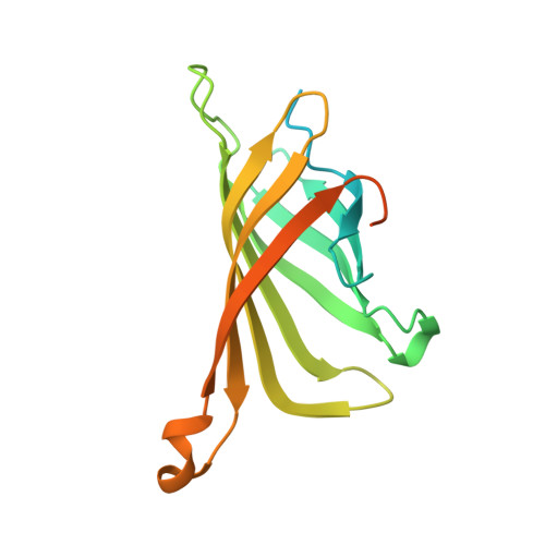
    Considerable efforts have been made to develop technologies for selection of peptidic molecules that act as substrates or binders to a protein of interest. Here we demonstrate the combination of rational peptide array library design, parallel screening and stepwise evolution, to discover novel peptide hotspots. These hotspots can be systematically evolved to create high-affinity, high-specificity binding peptides to a protein target in a reproducible and digitally controlled process. The method can be applied to synthesize both linear and cyclic peptides, as well as peptides composed of natural and non-natural amino acid analogs, thereby enabling screens in a much diverse chemical space. We apply this method to stepwise evolve peptide binders to streptavidin, a protein studied for over two decades and report novel peptides that mimic key interactions of biotin to streptavidin.


  • Organizational Affiliation

    Roche Madison, 500 S Rosa Rd, Madison, WI, 53719, USA.


Macromolecules
Find similar proteins by:  (by identity cutoff)  |  3D Structure
Entity ID: 1
MoleculeChains Sequence LengthOrganismDetailsImage
Streptavidin
A, B, E, F, H
A, B, E, F, H, K, M, O
183Streptomyces avidiniiMutation(s): 0 
UniProt
Find proteins for P22629 (Streptomyces avidinii)
Explore P22629 
Go to UniProtKB:  P22629
Entity Groups  
Sequence Clusters30% Identity50% Identity70% Identity90% Identity95% Identity100% Identity
UniProt GroupP22629
Sequence Annotations
Expand
  • Reference Sequence

Find similar proteins by:  Sequence   |   3D Structure  

Entity ID: 2
MoleculeChains Sequence LengthOrganismDetailsImage
GLU-TRP-VAL-HIS-PRO-GLN-PHE-GLU-GLN-LYS-ALA-LYS Peptide
C, D, G, I, J
C, D, G, I, J, L, N, P
12Homo sapiensMutation(s): 0 
Entity Groups  
Sequence Clusters30% Identity50% Identity70% Identity90% Identity95% Identity100% Identity
Sequence Annotations
Expand
  • Reference Sequence
Experimental Data & Validation

Experimental Data

  • Method: X-RAY DIFFRACTION
  • Resolution: 1.12 Å
  • R-Value Free: 0.186 
  • R-Value Work: 0.160 
  • R-Value Observed: 0.161 
  • Space Group: P 1
Unit Cell:
Length ( Å )Angle ( ˚ )
a = 57.913α = 96.31
b = 64.806β = 97.15
c = 81.156γ = 84.09
Software Package:
Software NamePurpose
REFMACrefinement
XDSdata reduction
SADABSdata scaling
PHASERphasing

Structure Validation

View Full Validation Report


Entry History 

Deposition Data

Revision History  (Full details and data files)

  • Version 1.0: 2017-10-04
    Type: Initial release