5N2S

Crystal structure of stabilized A1 receptor in complex with PSB36 at 3.3A resolution


Experimental Data Snapshot

  • Method: X-RAY DIFFRACTION
  • Resolution: 3.30 Å
  • R-Value Free: 0.278 
  • R-Value Work: 0.256 
  • R-Value Observed: 0.257 

wwPDB Validation   3D Report Full Report


Ligand Structure Quality Assessment 


This is version 1.2 of the entry. See complete history


Literature

Structures of Human A1 and A2A Adenosine Receptors with Xanthines Reveal Determinants of Selectivity.

Cheng, R.K.Y.Segala, E.Robertson, N.Deflorian, F.Dore, A.S.Errey, J.C.Fiez-Vandal, C.Marshall, F.H.Cooke, R.M.

(2017) Structure 25: 1275-1285.e4

  • DOI: https://doi.org/10.1016/j.str.2017.06.012
  • Primary Citation of Related Structures:  
    5MZJ, 5MZP, 5N2R, 5N2S

  • PubMed Abstract: 

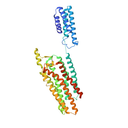
    The adenosine A 1 and A 2A receptors belong to the purinergic family of G protein-coupled receptors, and regulate diverse functions of the cardiovascular, respiratory, renal, inflammation, and CNS. Xanthines such as caffeine and theophylline are weak, non-selective antagonists of adenosine receptors. Here we report the structure of a thermostabilized human A 1 receptor at 3.3 Å resolution with PSB36, an A 1 -selective xanthine-based antagonist. This is compared with structures of the A 2A receptor with PSB36 (2.8 Å resolution), caffeine (2.1 Å), and theophylline (2.0 Å) to highlight features of ligand recognition which are common across xanthines. The structures of A 1 R and A 2A R were analyzed to identify the differences that are important selectivity determinants for xanthine ligands, and the role of T270 7.35 in A 1 R (M270 7.35 in A 2A R) in conferring selectivity was confirmed by mutagenesis. The structural differences confirmed to lead to selectivity can be utilized in the design of new subtype-selective A 1 R or A 2A R antagonists.


  • Organizational Affiliation

    Heptares Therapeutics Ltd, Biopark, Broadwater Road, Welwyn Garden City AL7 3AX, UK.


Macromolecules
Find similar proteins by:  (by identity cutoff)  |  3D Structure
Entity ID: 1
MoleculeChains Sequence LengthOrganismDetailsImage
Soluble cytochrome b562,Adenosine receptor A1436Escherichia coliHomo sapiens
This entity is chimeric
Mutation(s): 11 
Gene Names: cybCADORA1
Membrane Entity: Yes 
UniProt & NIH Common Fund Data Resources
Find proteins for P0ABE7 (Escherichia coli)
Explore P0ABE7 
Go to UniProtKB:  P0ABE7
Find proteins for P30542 (Homo sapiens)
Explore P30542 
Go to UniProtKB:  P30542
PHAROS:  P30542
GTEx:  ENSG00000163485 
Entity Groups  
Sequence Clusters30% Identity50% Identity70% Identity90% Identity95% Identity100% Identity
UniProt GroupsP30542P0ABE7
Sequence Annotations
Expand
  • Reference Sequence
Small Molecules
Ligands 2 Unique
IDChains Name / Formula / InChI Key2D Diagram3D Interactions
8K8
Query on 8K8

Download Ideal Coordinates CCD File 
B [auth A]1-butyl-3-(3-oxidanylpropyl)-8-[(1~{R},5~{S})-3-tricyclo[3.3.1.0^{3,7}]nonanyl]-7~{H}-purine-2,6-dione
C21 H30 N4 O3
CIBIXJYFYPFMTN-FZUGUKJMSA-N
SO4
Query on SO4

Download Ideal Coordinates CCD File 
C [auth A],
D [auth A]
SULFATE ION
O4 S
QAOWNCQODCNURD-UHFFFAOYSA-L
Experimental Data & Validation

Experimental Data

  • Method: X-RAY DIFFRACTION
  • Resolution: 3.30 Å
  • R-Value Free: 0.278 
  • R-Value Work: 0.256 
  • R-Value Observed: 0.257 
  • Space Group: C 2 2 2
Unit Cell:
Length ( Å )Angle ( ˚ )
a = 70.273α = 90
b = 111.658β = 90
c = 160.543γ = 90
Software Package:
Software NamePurpose
PHENIXrefinement
XDSdata reduction
Aimlessdata scaling
PHASERphasing

Structure Validation

View Full Validation Report


Ligand Structure Quality Assessment 


Entry History 

Deposition Data

Revision History  (Full details and data files)

  • Version 1.0: 2017-07-26
    Type: Initial release
  • Version 1.1: 2017-08-09
    Changes: Database references
  • Version 1.2: 2024-01-17
    Changes: Data collection, Database references, Refinement description