5MZE

Crystal structure of mouse MTH1 with 8-oxo-dGTP


Experimental Data Snapshot

  • Method: X-RAY DIFFRACTION
  • Resolution: 2.10 Å
  • R-Value Free: 0.239 
  • R-Value Work: 0.201 
  • R-Value Observed: 0.203 

wwPDB Validation   3D Report Full Report


Ligand Structure Quality Assessment 


This is version 1.4 of the entry. See complete history


Literature

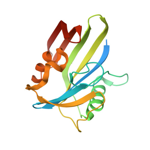
Crystal Structures and Inhibitor Interactions of Mouse and Dog MTH1 Reveal Species-Specific Differences in Affinity.

Narwal, M.Jemth, A.S.Gustafsson, R.Almlof, I.Warpman Berglund, U.Helleday, T.Stenmark, P.

(2018) Biochemistry 57: 593-603

  • DOI: https://doi.org/10.1021/acs.biochem.7b01163
  • Primary Citation of Related Structures:  
    5MZE, 5MZF, 5MZG, 6EHH

  • PubMed Abstract: 

    MTH1 hydrolyzes oxidized nucleoside triphosphates, thereby sanitizing the nucleotide pool from oxidative damage. This prevents incorporation of damaged nucleotides into DNA, which otherwise would lead to mutations and cell death. The high level of reactive oxygen species in cancer cells leads to a higher level of oxidized nucleotides in cancer cells compared to that in nonmalignant cells, making cancer cells more dependent on MTH1 for survival. The possibility of specifically targeting cancer cells by inhibiting MTH1 has highlighted MTH1 as a promising cancer target. The progression of MTH1 inhibitors into the clinic requires animal studies, and knowledge of species differences in the potency of inhibitors is vitally important. We here show that the human MTH1 inhibitor TH588 is approximately 20-fold less potent with respect to inhibition of mouse MTH1 than the human, rat, pig, and dog MTH1 proteins are. We present the crystal structures of mouse MTH1 in complex with TH588 and dog MTH1 and elucidate the structural and sequence basis for the observed difference in affinity for TH588. We identify amino acid residue 116 in MTH1 as an important determinant of TH588 affinity. Furthermore, we present the structure of mouse MTH1 in complex with the substrate 8-oxo-dGTP. The crystal structures provide insight into the high degree of structural conservation between MTH1 proteins from different organisms and provide a detailed view of interactions between MTH1 and the inhibitor, revealing that minute structural differences can have a large impact on affinity and specificity.


  • Organizational Affiliation

    Department of Biochemistry and Biophysics, Stockholm University , S-106 91 Stockholm, Sweden.


Macromolecules
Find similar proteins by:  (by identity cutoff)  |  3D Structure
Entity ID: 1
MoleculeChains Sequence LengthOrganismDetailsImage
7,8-dihydro-8-oxoguanine triphosphatase
A, B, C, D
176Mus musculusMutation(s): 0 
Gene Names: Nudt1Mth1
EC: 3.6.1.55 (PDB Primary Data), 3.6.1.56 (PDB Primary Data)
UniProt
Find proteins for P53368 (Mus musculus)
Explore P53368 
Go to UniProtKB:  P53368
Entity Groups  
Sequence Clusters30% Identity50% Identity70% Identity90% Identity95% Identity100% Identity
UniProt GroupP53368
Sequence Annotations
Expand
  • Reference Sequence
Small Molecules
Ligands 4 Unique
IDChains Name / Formula / InChI Key2D Diagram3D Interactions
8DG
Query on 8DG

Download Ideal Coordinates CCD File 
H [auth A],
K [auth B],
M [auth C],
O [auth D]
8-OXO-2'-DEOXYGUANOSINE-5'-TRIPHOSPHATE
C10 H16 N5 O14 P3
BUZOGVVQWCXXDP-VPENINKCSA-N
PEG
Query on PEG

Download Ideal Coordinates CCD File 
G [auth A],
N [auth D]
DI(HYDROXYETHYL)ETHER
C4 H10 O3
MTHSVFCYNBDYFN-UHFFFAOYSA-N
GOL
Query on GOL

Download Ideal Coordinates CCD File 
J [auth B]GLYCEROL
C3 H8 O3
PEDCQBHIVMGVHV-UHFFFAOYSA-N
CU
Query on CU

Download Ideal Coordinates CCD File 
E [auth A],
F [auth A],
I [auth B],
L [auth C]
COPPER (II) ION
Cu
JPVYNHNXODAKFH-UHFFFAOYSA-N
Experimental Data & Validation

Experimental Data

  • Method: X-RAY DIFFRACTION
  • Resolution: 2.10 Å
  • R-Value Free: 0.239 
  • R-Value Work: 0.201 
  • R-Value Observed: 0.203 
  • Space Group: P 1 21 1
Unit Cell:
Length ( Å )Angle ( ˚ )
a = 58.532α = 90
b = 139.245β = 107.53
c = 58.718γ = 90
Software Package:
Software NamePurpose
REFMACrefinement
XDSdata reduction
Aimlessdata scaling
PHASERphasing

Structure Validation

View Full Validation Report


Ligand Structure Quality Assessment 


Entry History 

Deposition Data

Revision History  (Full details and data files)

  • Version 1.0: 2018-01-10
    Type: Initial release
  • Version 1.1: 2018-01-24
    Changes: Database references
  • Version 1.2: 2018-02-14
    Changes: Database references
  • Version 1.3: 2018-11-07
    Changes: Data collection, Source and taxonomy
  • Version 1.4: 2024-01-17
    Changes: Data collection, Database references, Derived calculations, Refinement description