5MZ6

Cryo-EM structure of a Separase-Securin complex from Caenorhabditis elegans at 3.8 A resolution


Experimental Data Snapshot

  • Method: ELECTRON MICROSCOPY
  • Resolution: 3.80 Å
  • Aggregation State: PARTICLE 
  • Reconstruction Method: SINGLE PARTICLE 

wwPDB Validation   3D Report Full Report


This is version 1.4 of the entry. See complete history


Literature

Cryo-EM structure of a metazoan separase-securin complex at near-atomic resolution.

Boland, A.Martin, T.G.Zhang, Z.Yang, J.Bai, X.C.Chang, L.Scheres, S.H.Barford, D.

(2017) Nat Struct Mol Biol 24: 414-418

  • DOI: https://doi.org/10.1038/nsmb.3386
  • Primary Citation of Related Structures:  
    5MZ6

  • PubMed Abstract: 

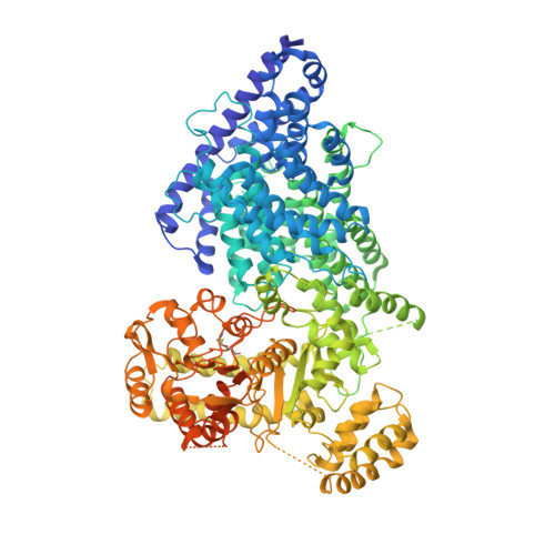
    Separase is a caspase-family protease that initiates chromatid segregation by cleaving the kleisin subunits (Scc1 and Rec8) of cohesin, and regulates centrosome duplication and mitotic spindle function through cleavage of kendrin and Slk19. To understand the mechanisms of securin regulation of separase, we used single-particle cryo-electron microscopy (cryo-EM) to determine a near-atomic-resolution structure of the Caenorhabditis elegans separase-securin complex. Separase adopts a triangular-shaped bilobal architecture comprising an N-terminal tetratricopeptide repeat (TPR)-like α-solenoid domain docked onto the conserved C-terminal protease domain. Securin engages separase in an extended antiparallel conformation, interacting with both lobes. It inhibits separase by interacting with the catalytic site through a pseudosubstrate mechanism, thus revealing that in the inhibited separase-securin complex, the catalytic site adopts a conformation compatible with substrate binding. Securin is protected from cleavage because an aliphatic side chain at the P1 position represses protease activity by disrupting the organization of catalytic site residues.


  • Organizational Affiliation

    MRC Laboratory of Molecular Biology, Cambridge, UK.


Macromolecules
Find similar proteins by:  (by identity cutoff)  |  3D Structure
Entity ID: 1
MoleculeChains Sequence LengthOrganismDetailsImage
SEParaseA [auth 1]1,262Caenorhabditis elegansMutation(s): 0 
Gene Names: sep-1CELE_Y47G6A.12Y47G6A.12
UniProt
Find proteins for G5ED39 (Caenorhabditis elegans)
Explore G5ED39 
Go to UniProtKB:  G5ED39
Entity Groups  
Sequence Clusters30% Identity50% Identity70% Identity90% Identity95% Identity100% Identity
UniProt GroupG5ED39
Sequence Annotations
Expand
  • Reference Sequence
Find similar proteins by:  (by identity cutoff)  |  3D Structure
Entity ID: 2
MoleculeChains Sequence LengthOrganismDetailsImage
Interactor of FizzY protein244Caenorhabditis elegansMutation(s): 0 
Gene Names: ify-1C27A2.3CELE_C27A2.3
UniProt
Find proteins for Q18235 (Caenorhabditis elegans)
Explore Q18235 
Go to UniProtKB:  Q18235
Entity Groups  
Sequence Clusters30% Identity50% Identity70% Identity90% Identity95% Identity100% Identity
UniProt GroupQ18235
Sequence Annotations
Expand
  • Reference Sequence
Experimental Data & Validation

Experimental Data

  • Method: ELECTRON MICROSCOPY
  • Resolution: 3.80 Å
  • Aggregation State: PARTICLE 
  • Reconstruction Method: SINGLE PARTICLE 
EM Software:
TaskSoftware PackageVersion
RECONSTRUCTIONRELION2.0

Structure Validation

View Full Validation Report


Entry History 

Deposition Data

Revision History  (Full details and data files)

  • Version 1.0: 2017-03-08
    Type: Initial release
  • Version 1.1: 2017-03-15
    Changes: Database references
  • Version 1.2: 2017-04-19
    Changes: Database references
  • Version 1.3: 2017-08-30
    Changes: Data collection
  • Version 1.4: 2019-12-11
    Changes: Other