5MYV

Crystal structure of SRPK2 in complex with compound 1


Experimental Data Snapshot

  • Method: X-RAY DIFFRACTION
  • Resolution: 2.90 Å
  • R-Value Free: 0.217 
  • R-Value Work: 0.199 
  • R-Value Observed: 0.200 

wwPDB Validation   3D Report Full Report


Ligand Structure Quality Assessment 


This is version 1.1 of the entry. See complete history


Literature

Development of Potent, Selective SRPK1 Inhibitors as Potential Topical Therapeutics for Neovascular Eye Disease.

Batson, J.Toop, H.D.Redondo, C.Babaei-Jadidi, R.Chaikuad, A.Wearmouth, S.F.Gibbons, B.Allen, C.Tallant, C.Zhang, J.Du, C.Hancox, J.C.Hawtrey, T.Da Rocha, J.Griffith, R.Knapp, S.Bates, D.O.Morris, J.C.

(2017) ACS Chem Biol 12: 825-832

  • DOI: https://doi.org/10.1021/acschembio.6b01048
  • Primary Citation of Related Structures:  
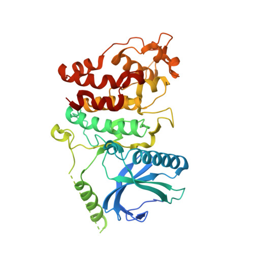
    5MXX, 5MY8, 5MYV

  • PubMed Abstract: 

    Serine/arginine-protein kinase 1 (SRPK1) regulates alternative splicing of VEGF-A to pro-angiogenic isoforms and SRPK1 inhibition can restore the balance of pro/antiangiogenic isoforms to normal physiological levels. The lack of potency and selectivity of available compounds has limited development of SRPK1 inhibitors, with the control of alternative splicing by splicing factor-specific kinases yet to be translated. We present here compounds that occupy a binding pocket created by the unique helical insert of SRPK1, and trigger a backbone flip in the hinge region, that results in potent (<10 nM) and selective inhibition of SRPK1 kinase activity. Treatment with these inhibitors inhibited SRPK1 activity and phosphorylation of serine/arginine splicing factor 1 (SRSF1), resulting in alternative splicing of VEGF-A from pro-angiogenic to antiangiogenic isoforms. This property resulted in potent inhibition of blood vessel growth in models of choroidal angiogenesis in vivo. This work identifies tool compounds for splice isoform selective targeting of pro-angiogenic VEGF, which may lead to new therapeutic strategies for a diversity of diseases where dysfunctional splicing drives disease development.


  • Organizational Affiliation

    Exonate Ltd , Unit 23, Cambridge Science Park, Cambridge, United Kingdom.


Macromolecules
Find similar proteins by:  (by identity cutoff)  |  3D Structure
Entity ID: 1
MoleculeChains Sequence LengthOrganismDetailsImage
SRSF protein kinase 2,SRSF protein kinase 2
A, B, C, D
389Homo sapiensMutation(s): 0 
Gene Names: SRPK2
EC: 2.7.11.1
UniProt & NIH Common Fund Data Resources
Find proteins for P78362 (Homo sapiens)
Explore P78362 
Go to UniProtKB:  P78362
PHAROS:  P78362
GTEx:  ENSG00000135250 
Entity Groups  
Sequence Clusters30% Identity50% Identity70% Identity90% Identity95% Identity100% Identity
UniProt GroupP78362
Sequence Annotations
Expand
  • Reference Sequence
Small Molecules
Ligands 3 Unique
IDChains Name / Formula / InChI Key2D Diagram3D Interactions
W4A
Query on W4A

Download Ideal Coordinates CCD File 
FB [auth D],
JA [auth B],
T [auth A],
UA [auth C]
5-methyl-~{N}-[2-(4-methylpiperazin-1-yl)-5-(trifluoromethyl)phenyl]furan-2-carboxamide
C18 H20 F3 N3 O2
STWLHUZLPCVTAS-UHFFFAOYSA-N
SO4
Query on SO4

Download Ideal Coordinates CCD File 
AA [auth B]
AB [auth D]
BA [auth B]
BB [auth D]
CA [auth B]
AA [auth B],
AB [auth D],
BA [auth B],
BB [auth D],
CA [auth B],
CB [auth D],
DA [auth B],
DB [auth D],
E [auth A],
EA [auth B],
EB [auth D],
F [auth A],
FA [auth B],
G [auth A],
GA [auth B],
H [auth A],
HA [auth B],
I [auth A],
IA [auth B],
J [auth A],
K [auth A],
KA [auth C],
L [auth A],
LA [auth C],
M [auth A],
MA [auth C],
N [auth A],
NA [auth C],
O [auth A],
OA [auth C],
P [auth A],
PA [auth C],
Q [auth A],
QA [auth C],
R [auth A],
RA [auth C],
S [auth A],
SA [auth C],
TA [auth C],
V [auth B],
W [auth B],
WA [auth D],
X [auth B],
XA [auth D],
Y [auth B],
YA [auth D],
Z [auth B],
ZA [auth D]
SULFATE ION
O4 S
QAOWNCQODCNURD-UHFFFAOYSA-L
DMS
Query on DMS

Download Ideal Coordinates CCD File 
GB [auth D],
U [auth A],
VA [auth C]
DIMETHYL SULFOXIDE
C2 H6 O S
IAZDPXIOMUYVGZ-UHFFFAOYSA-N
Experimental Data & Validation

Experimental Data

  • Method: X-RAY DIFFRACTION
  • Resolution: 2.90 Å
  • R-Value Free: 0.217 
  • R-Value Work: 0.199 
  • R-Value Observed: 0.200 
  • Space Group: C 2 2 21
Unit Cell:
Length ( Å )Angle ( ˚ )
a = 188.79α = 90
b = 248.99β = 90
c = 146.73γ = 90
Software Package:
Software NamePurpose
REFMACrefinement
XDSdata reduction
Aimlessdata scaling
PHASERphasing

Structure Validation

View Full Validation Report


Ligand Structure Quality Assessment 


Entry History 

Revision History  (Full details and data files)

  • Version 1.0: 2017-05-10
    Type: Initial release
  • Version 1.1: 2024-01-17
    Changes: Data collection, Database references, Refinement description