5MU5

Structure of MAf glycosyltransferase from Magnetospirillum magneticum AMB-1


Experimental Data Snapshot

  • Method: X-RAY DIFFRACTION
  • Resolution: 2.30 Å
  • R-Value Free: 0.220 
  • R-Value Work: 0.187 
  • R-Value Observed: 0.188 

wwPDB Validation   3D Report Full Report


This is version 1.2 of the entry. See complete history


Literature

Glycosylate and move! The glycosyltransferase Maf is involved in bacterial flagella formation.

Sulzenbacher, G.Roig-Zamboni, V.Lebrun, R.Guerardel, Y.Murat, D.Mansuelle, P.Yamakawa, N.Qian, X.X.Vincentelli, R.Bourne, Y.Wu, L.F.Alberto, F.

(2018) Environ Microbiol 20: 228-240

  • DOI: https://doi.org/10.1111/1462-2920.13975
  • Primary Citation of Related Structures:  
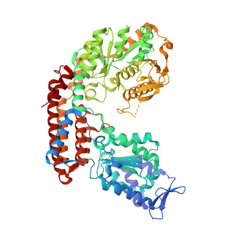
    5MU5

  • PubMed Abstract: 

    The flagella of various Gram-negative bacteria are decorated with diverse glycan structures, amongst them nonulosonic acids related to the sialic acid family. Although nonulosonic sugar biosynthesis pathways have been dissected in various pathogens, the enzymes transferring the sugars onto flagellin are still poorly characterized. The deletion of genes coding for motility associated factors (Mafs) found in many pathogenic strains systematically gives rise to nonflagellated bacteria lacking specific nonulosonic sugars on the flagellins, therefore, relating Maf function to flagellin glycosylation and bacterial motility. We investigated the role of Maf from our model organism, Magnetospirillum magneticum AMB-1, in the glycosylation and formation of the flagellum. Deletion of the gene amb0685 coding for Maf produced a nonflagellated bacterium where the flagellin was still produced but no longer glycosylated. Our X-ray structure analysis revealed that the central domain of Maf exhibits similarity to sialyltransferases from Campylobacter jejuni. Glycan analysis suggested that the nonulosonic carbohydrate structure transferred is pseudaminic acid or a very close derivative. This work describes the importance of glycosylation in the formation of the bacterial flagellum and provides the first structural model for a member of a new bacterial glycosyltransferase family involved in nonulosonic acids transfer onto flagellins.


  • Organizational Affiliation

    Aix Marseille Univ, CNRS, AFMB UMR7257, Marseille 13288, France.


Macromolecules
Find similar proteins by:  (by identity cutoff)  |  3D Structure
Entity ID: 1
MoleculeChains Sequence LengthOrganismDetailsImage
Uncharacterized protein666Paramagnetospirillum magneticum AMB-1Mutation(s): 0 
Gene Names: amb0685
UniProt
Find proteins for Q2W9I6 (Magnetospirillum magneticum (strain AMB-1 / ATCC 700264))
Explore Q2W9I6 
Go to UniProtKB:  Q2W9I6
Entity Groups  
Sequence Clusters30% Identity50% Identity70% Identity90% Identity95% Identity100% Identity
UniProt GroupQ2W9I6
Sequence Annotations
Expand
  • Reference Sequence
Small Molecules
Ligands 5 Unique
IDChains Name / Formula / InChI Key2D Diagram3D Interactions
PG4
Query on PG4

Download Ideal Coordinates CCD File 
F [auth A]TETRAETHYLENE GLYCOL
C8 H18 O5
UWHCKJMYHZGTIT-UHFFFAOYSA-N
PGE
Query on PGE

Download Ideal Coordinates CCD File 
E [auth A]TRIETHYLENE GLYCOL
C6 H14 O4
ZIBGPFATKBEMQZ-UHFFFAOYSA-N
SO4
Query on SO4

Download Ideal Coordinates CCD File 
B [auth A]SULFATE ION
O4 S
QAOWNCQODCNURD-UHFFFAOYSA-L
EDO
Query on EDO

Download Ideal Coordinates CCD File 
G [auth A],
H [auth A],
I [auth A],
J [auth A]
1,2-ETHANEDIOL
C2 H6 O2
LYCAIKOWRPUZTN-UHFFFAOYSA-N
CL
Query on CL

Download Ideal Coordinates CCD File 
C [auth A],
D [auth A]
CHLORIDE ION
Cl
VEXZGXHMUGYJMC-UHFFFAOYSA-M
Experimental Data & Validation

Experimental Data

  • Method: X-RAY DIFFRACTION
  • Resolution: 2.30 Å
  • R-Value Free: 0.220 
  • R-Value Work: 0.187 
  • R-Value Observed: 0.188 
  • Space Group: P 21 21 2
Unit Cell:
Length ( Å )Angle ( ˚ )
a = 117.447α = 90
b = 126.852β = 90
c = 64.601γ = 90
Software Package:
Software NamePurpose
REFMACrefinement
XDSdata reduction
SCALAdata scaling
CRANKphasing

Structure Validation

View Full Validation Report


Entry History 

Deposition Data

Revision History  (Full details and data files)

  • Version 1.0: 2017-11-15
    Type: Initial release
  • Version 1.1: 2018-01-24
    Changes: Database references
  • Version 1.2: 2019-04-10
    Changes: Data collection, Source and taxonomy