5MSK

Mouse PA28beta


Experimental Data Snapshot

  • Method: X-RAY DIFFRACTION
  • Resolution: 3.60 Å
  • R-Value Free: 0.263 
  • R-Value Work: 0.242 
  • R-Value Observed: 0.243 

wwPDB Validation   3D Report Full Report


This is version 1.2 of the entry. See complete history


Literature

The Mammalian Proteasome Activator PA28 Forms an Asymmetric alpha 4 beta 3 Complex.

Huber, E.M.Groll, M.

(2017) Structure 25: 1473-1480.e3

  • DOI: https://doi.org/10.1016/j.str.2017.07.013
  • Primary Citation of Related Structures:  
    5MSJ, 5MSK, 5MX5

  • PubMed Abstract: 

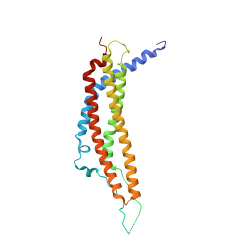
    The heptameric proteasome activator (PA) 28αβ is known to modulate class I antigen processing by docking onto 20S proteasome core particles (CPs). The exact stoichiometry and arrangement of its α and β subunits, however, is still controversial. Here we analyzed murine PA28 complexes regarding structure and assembly. Strikingly, PA28α, PA28β, and PA28αβ preparations form heptamers, but solely PA28α and PA28αβ associate with CPs. Co-expression of α and β yields one unique PA28αβ species with an unchangeable subunit composition. Structural data on PA28α, PA28β, and PA28αβ up to 2.9 Å resolution reveal a PA28α 4 β 3 complex with an alternating subunit arrangement and a single α-α interface. Differential scanning fluorimetry experiments and activity assays classify PA28α 4 β 3 as most stable and most active, indicating that this assembly might represent the physiologically relevant species. Together, our data resolve subunit composition and arrangement of PA28αβ and clarify how an asymmetric heptamer can be assembled from two highly homologous subunits.


  • Organizational Affiliation

    Center for Integrated Protein Science, Department Chemie, Lehrstuhl für Biochemie, Technische Universität München, Lichtenbergstrasse 4, 85748 Garching, Germany. Electronic address: eva.huber@tum.de.


Macromolecules
Find similar proteins by:  (by identity cutoff)  |  3D Structure
Entity ID: 1
MoleculeChains Sequence LengthOrganismDetailsImage
Proteasome activator complex subunit 2
A, B, C, D, E
A, B, C, D, E, F, G
239Mus musculusMutation(s): 0 
Gene Names: Psme2Pa28b1
UniProt
Find proteins for P97372 (Mus musculus)
Explore P97372 
Go to UniProtKB:  P97372
Entity Groups  
Sequence Clusters30% Identity50% Identity70% Identity90% Identity95% Identity100% Identity
UniProt GroupP97372
Sequence Annotations
Expand
  • Reference Sequence
Experimental Data & Validation

Experimental Data

  • Method: X-RAY DIFFRACTION
  • Resolution: 3.60 Å
  • R-Value Free: 0.263 
  • R-Value Work: 0.242 
  • R-Value Observed: 0.243 
  • Space Group: P 1 21 1
Unit Cell:
Length ( Å )Angle ( ˚ )
a = 86.84α = 90
b = 132.61β = 92.19
c = 90.93γ = 90
Software Package:
Software NamePurpose
REFMACrefinement
XDSdata reduction
XSCALEdata scaling
PHASERphasing

Structure Validation

View Full Validation Report


Entry History & Funding Information

Deposition Data


Funding OrganizationLocationGrant Number
German Research FoundationGermanySFB1035

Revision History  (Full details and data files)

  • Version 1.0: 2017-09-13
    Type: Initial release
  • Version 1.1: 2017-10-18
    Changes: Database references
  • Version 1.2: 2024-01-17
    Changes: Data collection, Database references, Refinement description