Intact Protein Analysis at 21 Tesla and X-Ray Crystallography Define Structural Differences in Single Amino Acid Variants of Human Mitochondrial Branched-Chain Amino Acid Aminotransferase 2 (BCAT2).
Anderson, L.C., Hakansson, M., Walse, B., Nilsson, C.L.(2017) J Am Soc Mass Spectrom 28: 1796-1804
- PubMed: 28681360
- DOI: https://doi.org/10.1007/s13361-017-1705-0
- Primary Citation of Related Structures:
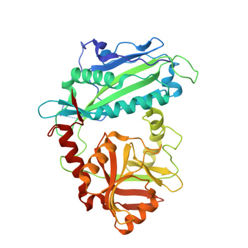
5MPR - PubMed Abstract:
Structural technologies are an essential component in the design of precision therapeutics. Precision medicine entails the development of therapeutics directed toward a designated target protein, with the goal to deliver the right drug to the right patient at the right time. In the field of oncology, protein structural variants are often associated with oncogenic potential. In a previous proteogenomic screen of patient-derived glioblastoma (GBM) tumor materials, we identified a sequence variant of human mitochondrial branched-chain amino acid aminotransferase 2 as a putative factor of resistance of GBM to standard-of-care-treatments. The enzyme generates glutamate, which is neurotoxic. To elucidate structural coordinates that may confer altered substrate binding or activity of the variant BCAT2 T186R, a ~45 kDa protein, we applied combined ETD and CID top-down mass spectrometry in a LC-FT-ICR MS at 21 T, and X-Ray crystallography in the study of both the variant and non-variant intact proteins. The combined ETD/CID fragmentation pattern allowed for not only extensive sequence coverage but also confident localization of the amino acid variant to its position in the sequence. The crystallographic experiments confirmed the hypothesis generated by in silico structural homology modeling, that the Lys59 side-chain of BCAT2 may repulse the Arg186 in the variant protein (PDB code: 5MPR), leading to destabilization of the protein dimer and altered enzyme kinetics. Taken together, the MS and novel 3D structural data give us reason to further pursue BCAT2 T186R as a precision drug target in GBM. Graphical Abstract ᅟ.
Organizational Affiliation:
Ion Cyclotron Resonance Program, National High Magnetic Field Laboratory, 1800 E. Paul Dirac Dr., Tallahassee, FL, 32310, USA.