5MOC

Crystal structure of 14-3-3sigma and a p53 C-terminal 12-mer synthetic phosphopeptide


Experimental Data Snapshot

  • Method: X-RAY DIFFRACTION
  • Resolution: 1.80 Å
  • R-Value Free: 0.208 
  • R-Value Work: 0.165 
  • R-Value Observed: 0.167 

wwPDB Validation   3D Report Full Report


This is version 2.1 of the entry. See complete history


Literature

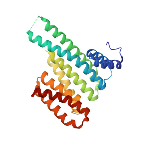
Small-molecule stabilization of the p53 - 14-3-3 protein-protein interaction.

Doveston, R.G.Kuusk, A.Andrei, S.A.Leysen, S.Cao, Q.Castaldi, M.P.Hendricks, A.Brunsveld, L.Chen, H.Boyd, H.Ottmann, C.

(2017) FEBS Lett 591: 2449-2457

  • DOI: https://doi.org/10.1002/1873-3468.12723
  • Primary Citation of Related Structures:  
    5MHC, 5MOC, 5MXO

  • PubMed Abstract: 

    14-3-3 proteins are positive regulators of the tumor suppressor p53, the mutation of which is implicated in many human cancers. Current strategies for targeting of p53 involve restoration of wild-type function or inhibition of the interaction with MDM2, its key negative regulator. Despite the efficacy of these strategies, the alternate approach of stabilizing the interaction of p53 with positive regulators and, thus, enhancing tumor suppressor activity, has not been explored. Here, we report the first example of small-molecule stabilization of the 14-3-3 - p53 protein-protein interaction (PPI) and demonstrate the potential of this approach as a therapeutic modality. We also observed a disconnect between biophysical and crystallographic data in the presence of a stabilizing molecule, which is unusual in 14-3-3 PPIs.


  • Organizational Affiliation

    Laboratory of Chemical Biology, Department of Biomedical Engineering and Institute for Complex Molecular Systems, Eindhoven University of Technology, The Netherlands.


Macromolecules
Find similar proteins by:  (by identity cutoff)  |  3D Structure
Entity ID: 1
MoleculeChains Sequence LengthOrganismDetailsImage
14-3-3 protein sigma236Homo sapiensMutation(s): 0 
Gene Names: SFNHME1
UniProt & NIH Common Fund Data Resources
Find proteins for P31947 (Homo sapiens)
Explore P31947 
Go to UniProtKB:  P31947
PHAROS:  P31947
GTEx:  ENSG00000175793 
Entity Groups  
Sequence Clusters30% Identity50% Identity70% Identity90% Identity95% Identity100% Identity
UniProt GroupP31947
Sequence Annotations
Expand
  • Reference Sequence

Find similar proteins by:  Sequence   |   3D Structure  

Entity ID: 2
MoleculeChains Sequence LengthOrganismDetailsImage
p53 C-terminal domainB [auth P]12Homo sapiensMutation(s): 0 
UniProt & NIH Common Fund Data Resources
Find proteins for P04637 (Homo sapiens)
Explore P04637 
Go to UniProtKB:  P04637
PHAROS:  P04637
GTEx:  ENSG00000141510 
Entity Groups  
Sequence Clusters30% Identity50% Identity70% Identity90% Identity95% Identity100% Identity
UniProt GroupP04637
Sequence Annotations
Expand
  • Reference Sequence
Small Molecules
Modified Residues  1 Unique
IDChains TypeFormula2D DiagramParent
TPO
Query on TPO
B [auth P]L-PEPTIDE LINKINGC4 H10 N O6 PTHR
Experimental Data & Validation

Experimental Data

  • Method: X-RAY DIFFRACTION
  • Resolution: 1.80 Å
  • R-Value Free: 0.208 
  • R-Value Work: 0.165 
  • R-Value Observed: 0.167 
  • Space Group: C 2 2 21
Unit Cell:
Length ( Å )Angle ( ˚ )
a = 81.91α = 90
b = 112.27β = 90
c = 62.42γ = 90
Software Package:
Software NamePurpose
Aimlessdata scaling
PHASERphasing
PHENIXrefinement
PDB_EXTRACTdata extraction
iMOSFLMdata processing

Structure Validation

View Full Validation Report


Entry History & Funding Information

Deposition Data


Funding OrganizationLocationGrant Number
Marie CurieNetherlandsPIAP-GA-2011-286418 14-3-3stab

Revision History  (Full details and data files)

  • Version 1.0: 2017-10-04
    Type: Initial release
  • Version 2.0: 2019-03-13
    Changes: Atomic model, Data collection, Derived calculations
  • Version 2.1: 2024-01-17
    Changes: Data collection, Database references, Refinement description