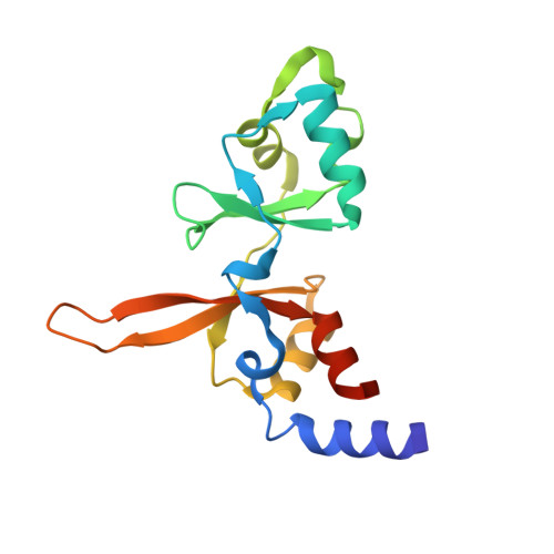
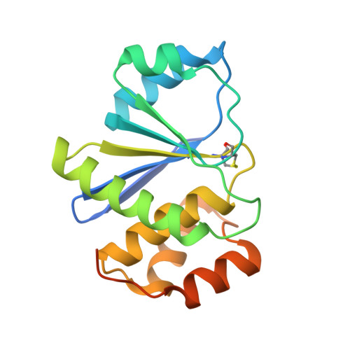
Structural Basis of the Oncogenic Interaction of Phosphatase PRL-1 with the Magnesium Transporter CNNM2.
Gimenez-Mascarell, P., Oyenarte, I., Hardy, S., Breiderhoff, T., Stuiver, M., Kostantin, E., Diercks, T., Pey, A.L., Ereno-Orbea, J., Martinez-Chantar, M.L., Khalaf-Nazzal, R., Claverie-Martin, F., Muller, D., Tremblay, M.L., Martinez-Cruz, L.A.(2017) J Biol Chem 292: 786-801
- PubMed: 27899452
- DOI: https://doi.org/10.1074/jbc.M116.759944
- Primary Citation of Related Structures:
5LXQ, 5MMZ - PubMed Abstract:
Phosphatases of regenerating liver (PRLs), the most oncogenic of all protein-tyrosine phosphatases (PTPs), play a critical role in metastatic progression of cancers. Recent findings established a new paradigm by uncovering that their association with magnesium transporters of the cyclin M (CNNM) family causes a rise in intracellular magnesium levels that promote oncogenic transformation. Recently, however, essential roles for regulation of the circadian rhythm and reproduction of the CNNM family have been highlighted. Here, we describe the crystal structure of PRL-1 in complex with the Bateman module of CNNM2 (CNNM2 BAT ), which consists of two cystathionine β-synthase (CBS) domains (IPR000664) and represents an intracellular regulatory module of the transporter. The structure reveals a heterotetrameric association, consisting of a disc-like homodimer of CNNM2 BAT bound to two independent PRL-1 molecules, each one located at opposite tips of the disc. The structure highlights the key role played by Asp-558 at the extended loop of the CBS2 motif of CNNM2 in maintaining the association between the two proteins and proves that the interaction between CNNM2 and PRL-1 occurs via the catalytic domain of the phosphatase. Our data shed new light on the structural basis underlying the interaction between PRL phosphatases and CNNM transporters and provides a hypothesis about the molecular mechanism by which PRL-1, upon binding to CNNM2, might increase the intracellular concentration of Mg 2+ thereby contributing to tumor progression and metastasis. The availability of this structure sets the basis for the rational design of compounds modulating PRL-1 and CNNM2 activities.
Organizational Affiliation:
From the Structural Biology Unit, Center for Cooperative Research in Biosciences (CIC bioGUNE), Technology Park of Bizkaia, 48160 Derio, Bizkaia, Spain.