Muropeptide Binding and the X-ray Structure of the Effector Domain of the Transcriptional Regulator AmpR of Pseudomonas aeruginosa.
Dik, D.A., Dominguez-Gil, T., Lee, M., Hesek, D., Byun, B., Fishovitz, J., Boggess, B., Hellman, L.M., Fisher, J.F., Hermoso, J.A., Mobashery, S.(2017) J Am Chem Soc 139: 1448-1451
- PubMed: 28079369
- DOI: https://doi.org/10.1021/jacs.6b12819
- Primary Citation of Related Structures:
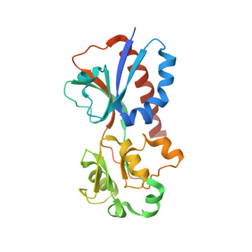
5MMH - PubMed Abstract:
A complex link exists between cell-wall recycling/repair and the manifestation of resistance to β-lactam antibiotics in many Enterobacteriaceae and Pseudomonas aeruginosa. This process is mediated by specific cell-wall-derived muropeptide products. These muropeptides are internalized into the cytoplasm and bind to the transcriptional regulator AmpR, which controls the cytoplasmic events that lead to expression of β-lactamase, an antibiotic-resistance determinant. The effector-binding domain (EBD) of AmpR was purified to homogeneity. We document that the EBD exists exclusively as a dimer, even at a concentration as low as 1 μM. The EBD binds to the suppressor ligand UDP-N-acetyl-β-d-muramyl-l-Ala-γ-d-Glu-meso-DAP-d-Ala-d-Ala and binds to two activator muropeptides, N-acetyl-β-d-glucosamine-(1→4)-1,6-anhydro-N-acetyl-β-d-muramyl-l-Ala-γ-d-Glu-meso-DAP-d-Ala-d-Ala and 1,6-anhydro-N-acetyl-β-d-muramyl-l-Ala-γ-d-Glu-meso-DAP-d-Ala-d-Ala, as assessed by non-denaturing mass spectrometry. The EBD does not bind to 1,6-anhydro-N-acetyl-β-d-muramyl-l-Ala-γ-d-Glu-meso-DAP. This binding selectivity revises the dogma in the field. The crystal structure of the EBD dimer was solved to 2.2 Å resolution. The EBD crystallizes in a "closed" conformation, in contrast to the "open" structure required to bind the muropeptides. Structural issues of this ligand recognition are addressed by molecular dynamics simulations, which reveal significant differences among the complexes with the effector molecules.
Organizational Affiliation:
Department of Chemistry and Biochemistry, University of Notre Dame , Notre Dame, Indiana 46556, United States.