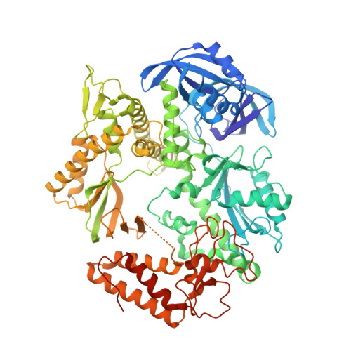
Structure of the family B DNA polymerase from the hyperthermophilic archaeon Pyrobaculum calidifontis.
Guo, J., Zhang, W., Coker, A.R., Wood, S.P., Cooper, J.B., Ahmad, S., Ali, S., Rashid, N., Akhtar, M.(2017) Acta Crystallogr D Struct Biol 73: 420-427
- PubMed: 28471366
- DOI: https://doi.org/10.1107/S2059798317004090
- Primary Citation of Related Structures:
5MDN - PubMed Abstract:
The family B DNA polymerase from Pyrobaculum calidifontis (Pc-polymerase) consists of 783 amino acids and is magnesium-ion dependent. It has an optimal pH of 8.5, an optimal temperature of 75°C and a half-life of 4.5 h at 95°C, giving it greater thermostability than the widely used Taq DNA polymerase. The enzyme is also capable of PCR-amplifying larger DNA fragments of up to 7.5 kb in length. It was shown to have functional, error-correcting 3'-5' exonuclease activity, as do the related high-fidelity DNA polymerases from Pyrococcus furiosus, Thermococcus kodakarensis KOD1 and Thermococcus gorgonarius, which have extensive commercial applications. Pc-polymerase has a quite low sequence identity of approximately 37% to these enzymes, which, in contrast, have very high sequence identity to each other, suggesting that the P. calidifontis enzyme is distinct. Here, the structure determination of Pc-polymerase is reported, which has been refined to an R factor of 24.47% and an R free of 28.81% at 2.80 Å resolution. The domains of the enzyme are arranged in a circular fashion to form a disc with a narrow central channel. One face of the disc has a number of connected crevices in it, which allow the protein to bind duplex and single-stranded DNA. The central channel is thought to allow incoming nucleoside triphosphates to access the active site. The enzyme has a number of unique structural features which distinguish it from other archaeal DNA polymerases and may account for its high processivity. A model of the complex with the primer-template duplex of DNA indicates that the largest conformational change that occurs upon DNA binding is the movement of the thumb domain, which rotates by 7.6° and moves by 10.0 Å. The surface potential of the enzyme is dominated by acidic groups in the central region of the molecule, where catalytic magnesium ions bind at the polymerase and exonuclease active sites. The outer regions are richer in basic amino acids that presumably interact with the sugar-phosphate backbone of DNA. The large number of salt bridges may contribute to the high thermal stability of this enzyme.
Organizational Affiliation:
Wolfson Institute for Biomedical Research, Division of Medicine, UCL, Gower Street, London WC1E 6BT, England.