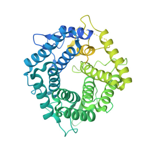
5M77

a GH76 family enzyme structure


Experimental Data Snapshot

  • Method: X-RAY DIFFRACTION
  • Resolution: 1.46 Å
  • R-Value Free: 0.165 
  • R-Value Work: 0.121 
  • R-Value Observed: 0.123 

wwPDB Validation   3D Report Full Report


This is version 2.1 of the entry. See complete history


Literature

An atypical interaction explains the high-affinity of a non-hydrolyzable S-linked 1,6-alpha-mannanase inhibitor.

Belz, T.Jin, Y.Coines, J.Rovira, C.Davies, G.J.Williams, S.J.

(2017) Chem Commun (Camb) 53: 9238-9241

  • DOI: https://doi.org/10.1039/c7cc04977c
  • Primary Citation of Related Structures:  
    5M77, 5N0F

  • PubMed Abstract: 

    The non-hydrolyzable S-linked azasugars, 1,6-α-mannosylthio- and 1,6-α-mannobiosylthioisofagomine, were synthesized and shown to bind with high affinity to a family 76 endo-1,6-α-mannanase from Bacillus circulans. X-ray crystallography showed an atypical interaction of the isofagomine nitrogen with the catalytic acid/base. Molecular dynamics simulations reveal that the atypical binding results from sulfur perturbing the most stable form away from the nucleophile interaction preferred for the O-linked congener.


  • Organizational Affiliation

    School of Chemistry and Bio21 Molecular Science and Biotechnology Institute, University of Melbourne, Parkville, Victoria 3010, Australia. sjwill@unimelb.edu.au.


Macromolecules
Find similar proteins by:  (by identity cutoff)  |  3D Structure
Entity ID: 1
MoleculeChains Sequence LengthOrganismDetailsImage
Alpha-1,6-mannanase
A, B
589Niallia circulansMutation(s): 1 
Gene Names: aman6
UniProt
Find proteins for Q9Z4P9 (Niallia circulans)
Explore Q9Z4P9 
Go to UniProtKB:  Q9Z4P9
Entity Groups  
Sequence Clusters30% Identity50% Identity70% Identity90% Identity95% Identity100% Identity
UniProt GroupQ9Z4P9
Sequence Annotations
Expand
  • Reference Sequence
Oligosaccharides

Help

Entity ID: 2
MoleculeChains Length2D Diagram Glycosylation3D Interactions
1-thio-alpha-D-mannopyranose-(1-6)-[(3S,4R,5R)-4,5-dihydroxypiperidin-3-yl]methyl 1-thio-alpha-D-mannopyranoside
C, D
2N/AN/A
Small Molecules
Ligands 1 Unique
IDChains Name / Formula / InChI Key2D Diagram3D Interactions
EDO
Query on EDO

Download Ideal Coordinates CCD File 
E [auth A]
F [auth A]
G [auth A]
H [auth A]
I [auth A]
E [auth A],
F [auth A],
G [auth A],
H [auth A],
I [auth A],
J [auth A],
K [auth B],
L [auth B],
M [auth B],
N [auth B],
O [auth B],
P [auth B],
Q [auth B],
R [auth B],
S [auth B]
1,2-ETHANEDIOL
C2 H6 O2
LYCAIKOWRPUZTN-UHFFFAOYSA-N
Experimental Data & Validation

Experimental Data

  • Method: X-RAY DIFFRACTION
  • Resolution: 1.46 Å
  • R-Value Free: 0.165 
  • R-Value Work: 0.121 
  • R-Value Observed: 0.123 
  • Space Group: P 21 21 21
Unit Cell:
Length ( Å )Angle ( ˚ )
a = 84.06α = 90
b = 85.17β = 90
c = 101.86γ = 90
Software Package:
Software NamePurpose
REFMACrefinement
xia2data reduction
xia2data scaling
MOLREPphasing

Structure Validation

View Full Validation Report


Entry History & Funding Information

Deposition Data


Funding OrganizationLocationGrant Number
European Research CouncilUnited KingdomAdG-322942

Revision History  (Full details and data files)

  • Version 1.0: 2017-08-09
    Type: Initial release
  • Version 1.1: 2017-08-16
    Changes: Database references
  • Version 1.2: 2017-08-23
    Changes: Database references
  • Version 2.0: 2020-07-29
    Type: Remediation
    Reason: Carbohydrate remediation
    Changes: Atomic model, Author supporting evidence, Data collection, Derived calculations, Structure summary
  • Version 2.1: 2024-01-17
    Changes: Data collection, Database references, Refinement description, Structure summary