5M73

Structure of the human SRP S domain with SRP72 RNA-binding domain


Experimental Data Snapshot

  • Method: X-RAY DIFFRACTION
  • Resolution: 3.40 Å
  • R-Value Free: 0.280 
  • R-Value Work: 0.237 
  • R-Value Observed: 0.239 

wwPDB Validation   3D Report Full Report


This is version 1.6 of the entry. See complete history


Literature

Structures of human SRP72 complexes provide insights into SRP RNA remodeling and ribosome interaction.

Becker, M.M.Lapouge, K.Segnitz, B.Wild, K.Sinning, I.

(2017) Nucleic Acids Res 45: 470-481

  • DOI: https://doi.org/10.1093/nar/gkw1124
  • Primary Citation of Related Structures:  
    5M72, 5M73

  • PubMed Abstract: 

    Co-translational protein targeting and membrane protein insertion is a fundamental process and depends on the signal recognition particle (SRP). In mammals, SRP is composed of the SRP RNA crucial for SRP assembly and function and six proteins. The two largest proteins SRP68 and SRP72 form a heterodimer and bind to a regulatory site of the SRP RNA. Despite their essential roles in the SRP pathway, structural information has been available only for the SRP68 RNA-binding domain (RBD). Here we present the crystal structures of the SRP68 protein-binding domain (PBD) in complex with SRP72-PBD and of the SRP72-RBD bound to the SRP S domain (SRP RNA, SRP19 and SRP68) detailing all interactions of SRP72 within SRP. The SRP72-PBD is a tetratricopeptide repeat, which binds an extended linear motif of SRP68 with high affinity. The SRP72-RBD is a flexible peptide crawling along the 5e- and 5f-loops of SRP RNA. A conserved tryptophan inserts into the 5e-loop forming a novel type of RNA kink-turn stabilized by a potassium ion, which we define as K + -turn. In addition, SRP72-RBD remodels the 5f-loop involved in ribosome binding and visualizes SRP RNA plasticity. Docking of the S domain structure into cryo-electron microscopy density maps reveals multiple contact sites between SRP68/72 and the ribosome, and explains the role of SRP72 in the SRP pathway.


  • Organizational Affiliation

    Heidelberg University Biochemistry Center (BZH), Im Neuenheimer Feld 328, D-69120 Heidelberg, Germany.


Macromolecules

Find similar proteins by:  (by identity cutoff)  |  3D Structure
Entity ID: 2
MoleculeChains Sequence LengthOrganismDetailsImage
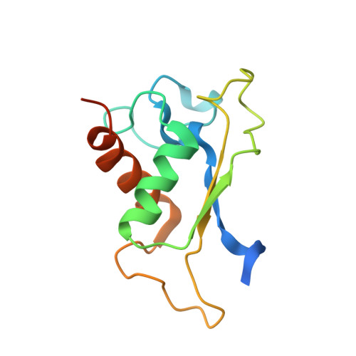
Signal recognition particle 19 kDa protein
B, F
128Homo sapiensMutation(s): 0 
Gene Names: SRP19
UniProt & NIH Common Fund Data Resources
Find proteins for P09132 (Homo sapiens)
Explore P09132 
Go to UniProtKB:  P09132
PHAROS:  P09132
GTEx:  ENSG00000153037 
Entity Groups  
Sequence Clusters30% Identity50% Identity70% Identity90% Identity95% Identity100% Identity
UniProt GroupP09132
Sequence Annotations
Expand
  • Reference Sequence
Find similar proteins by:  (by identity cutoff)  |  3D Structure
Entity ID: 3
MoleculeChains Sequence LengthOrganismDetailsImage
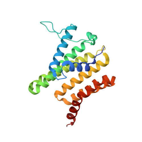
Signal recognition particle subunit SRP68
C, G
203Homo sapiensMutation(s): 0 
Gene Names: SRP68
UniProt & NIH Common Fund Data Resources
Find proteins for Q9UHB9 (Homo sapiens)
Explore Q9UHB9 
Go to UniProtKB:  Q9UHB9
PHAROS:  Q9UHB9
GTEx:  ENSG00000167881 
Entity Groups  
Sequence Clusters30% Identity50% Identity70% Identity90% Identity95% Identity100% Identity
UniProt GroupQ9UHB9
Sequence Annotations
Expand
  • Reference Sequence
Find similar proteins by:  (by identity cutoff)  |  3D Structure
Entity ID: 4
MoleculeChains Sequence LengthOrganismDetailsImage
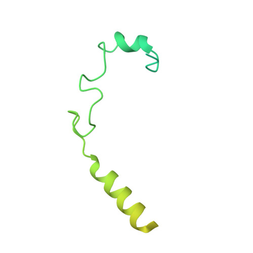
Signal recognition particle subunit SRP72
D, H
158Homo sapiensMutation(s): 0 
Gene Names: SRP72
UniProt & NIH Common Fund Data Resources
Find proteins for O76094 (Homo sapiens)
Explore O76094 
Go to UniProtKB:  O76094
PHAROS:  O76094
GTEx:  ENSG00000174780 
Entity Groups  
Sequence Clusters30% Identity50% Identity70% Identity90% Identity95% Identity100% Identity
UniProt GroupO76094
Sequence Annotations
Expand
  • Reference Sequence
Find similar nucleic acids by:  (by identity cutoff)  |  3D Structure
Entity ID: 1
MoleculeChains LengthOrganismImage
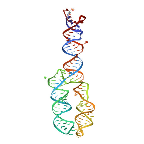
Human gene for small cytoplasmic 7SL RNA (7L30.1)
A, E
145Homo sapiens
Sequence Annotations
Expand
  • Reference Sequence
Small Molecules
Ligands 3 Unique
IDChains Name / Formula / InChI Key2D Diagram3D Interactions
GOL
Query on GOL

Download Ideal Coordinates CCD File 
OA [auth A]
PA [auth A]
QA [auth A]
QB [auth E]
RA [auth A]
OA [auth A],
PA [auth A],
QA [auth A],
QB [auth E],
RA [auth A],
RB [auth E],
SA [auth A],
SB [auth E],
TA [auth B]
GLYCEROL
C3 H8 O3
PEDCQBHIVMGVHV-UHFFFAOYSA-N
K
Query on K

Download Ideal Coordinates CCD File 
JA [auth A]
KA [auth A]
LA [auth A]
LB [auth E]
MA [auth A]
JA [auth A],
KA [auth A],
LA [auth A],
LB [auth E],
MA [auth A],
MB [auth E],
NA [auth A],
NB [auth E],
OB [auth E],
PB [auth E]
POTASSIUM ION
K
NPYPAHLBTDXSSS-UHFFFAOYSA-N
MG
Query on MG

Download Ideal Coordinates CCD File 
AA [auth A]
AB [auth E]
BA [auth A]
BB [auth E]
CA [auth A]
AA [auth A],
AB [auth E],
BA [auth A],
BB [auth E],
CA [auth A],
CB [auth E],
DA [auth A],
DB [auth E],
EA [auth A],
EB [auth E],
FA [auth A],
FB [auth E],
GA [auth A],
GB [auth E],
HA [auth A],
HB [auth E],
I [auth A],
IA [auth A],
IB [auth E],
J [auth A],
JB [auth E],
K [auth A],
KB [auth E],
L [auth A],
M [auth A],
N [auth A],
O [auth A],
P [auth A],
Q [auth A],
R [auth A],
S [auth A],
T [auth A],
TB [auth G],
U [auth A],
UA [auth C],
V [auth A],
VA [auth C],
W [auth A],
WA [auth C],
X [auth A],
XA [auth E],
Y [auth A],
YA [auth E],
Z [auth A],
ZA [auth E]
MAGNESIUM ION
Mg
JLVVSXFLKOJNIY-UHFFFAOYSA-N
Experimental Data & Validation

Experimental Data

  • Method: X-RAY DIFFRACTION
  • Resolution: 3.40 Å
  • R-Value Free: 0.280 
  • R-Value Work: 0.237 
  • R-Value Observed: 0.239 
  • Space Group: P 21 21 21
Unit Cell:
Length ( Å )Angle ( ˚ )
a = 134.567α = 90
b = 139.306β = 90
c = 152.246γ = 90
Software Package:
Software NamePurpose
PHENIXrefinement
PDB_EXTRACTdata extraction
XDSdata reduction
Aimlessdata scaling
PHASERphasing

Structure Validation

View Full Validation Report


Entry History & Funding Information

Deposition Data


Funding OrganizationLocationGrant Number
German Research FoundationGermanySFB 638
German Research FoundationGermanyGRK1188

Revision History  (Full details and data files)

  • Version 1.0: 2016-12-07
    Type: Initial release
  • Version 1.1: 2016-12-14
    Changes: Database references
  • Version 1.2: 2016-12-21
    Changes: Derived calculations
  • Version 1.3: 2017-01-18
    Changes: Database references
  • Version 1.4: 2019-10-16
    Changes: Author supporting evidence, Data collection, Derived calculations
  • Version 1.5: 2019-10-23
    Changes: Data collection, Database references
  • Version 1.6: 2024-01-17
    Changes: Data collection, Database references, Derived calculations, Refinement description