5M63

Crystal structure of group B Streptococcus type III DP2 oligosaccharide bound to Fab NVS-1-19-5


Experimental Data Snapshot

  • Method: X-RAY DIFFRACTION
  • Resolution: 2.74 Å
  • R-Value Free: 0.282 
  • R-Value Work: 0.236 
  • R-Value Observed: 0.238 

wwPDB Validation   3D Report Full Report


Ligand Structure Quality Assessment 


This is version 2.1 of the entry. See complete history


Literature

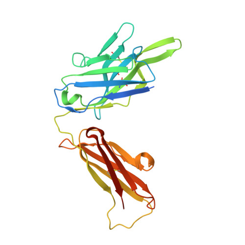
Structure of a protective epitope of group B Streptococcus type III capsular polysaccharide.

Carboni, F.Adamo, R.Fabbrini, M.De Ricco, R.Cattaneo, V.Brogioni, B.Veggi, D.Pinto, V.Passalacqua, I.Oldrini, D.Rappuoli, R.Malito, E.Margarit, I.Y.R.Berti, F.

(2017) Proc Natl Acad Sci U S A 114: 5017-5022

  • DOI: https://doi.org/10.1073/pnas.1701885114
  • Primary Citation of Related Structures:  
    5M63

  • PubMed Abstract: 

    Despite substantial progress in the prevention of group B Streptococcus (GBS) disease with the introduction of intrapartum antibiotic prophylaxis, this pathogen remains a leading cause of neonatal infection. Capsular polysaccharide conjugate vaccines have been tested in phase I/II clinical studies, showing promise for further development. Mapping of epitopes recognized by protective antibodies is crucial for understanding the mechanism of action of vaccines and for enabling antigen design. In this study, we report the structure of the epitope recognized by a monoclonal antibody with opsonophagocytic activity and representative of the protective response against type III GBS polysaccharide. The structure and the atomic-level interactions were determined by saturation transfer difference (STD)-NMR and X-ray crystallography using oligosaccharides obtained by synthetic and depolymerization procedures. The GBS PSIII epitope is made by six sugars. Four of them derive from two adjacent repeating units of the PSIII backbone and two of them from the branched galactose-sialic acid disaccharide contained in this sequence. The sialic acid residue establishes direct binding interactions with the functional antibody. The crystal structure provides insight into the molecular basis of antibody-carbohydrate interactions and confirms that the conformational epitope is not required for antigen recognition. Understanding the structural basis of immune recognition of capsular polysaccharide epitopes can aid in the design of novel glycoconjugate vaccines.


  • Organizational Affiliation

    GSK Vaccines, 53100 Siena, Italy.


Macromolecules
Find similar proteins by:  (by identity cutoff)  |  3D Structure
Entity ID: 1
MoleculeChains Sequence LengthOrganismDetailsImage
H chain of Fab NVS-1-19-5A [auth H],
C [auth M]
248Oryctolagus cuniculusMutation(s): 0 
Entity Groups  
Sequence Clusters30% Identity50% Identity70% Identity90% Identity95% Identity100% Identity
Sequence Annotations
Expand
  • Reference Sequence
Find similar proteins by:  (by identity cutoff)  |  3D Structure
Entity ID: 2
MoleculeChains Sequence LengthOrganismDetailsImage
L chain of Fab NVS-1-19-5B [auth L],
D [auth N]
239Oryctolagus cuniculusMutation(s): 0 
Entity Groups  
Sequence Clusters30% Identity50% Identity70% Identity90% Identity95% Identity100% Identity
Sequence Annotations
Expand
  • Reference Sequence
Oligosaccharides

Help

Entity ID: 3
MoleculeChains Length2D Diagram Glycosylation3D Interactions
N-acetyl-alpha-neuraminic acid-(2-3)-beta-D-galactopyranoseE [auth A],
G [auth C]
2N/A
Glycosylation Resources
GlyTouCan:  G30207PZ
GlyCosmos:  G30207PZ
GlyGen:  G30207PZ
Entity ID: 4
MoleculeChains Length2D Diagram Glycosylation3D Interactions
N-acetyl-alpha-neuraminic acid-(2-3)-beta-D-galactopyranose-(1-4)-[beta-D-galactopyranose-(1-4)-beta-D-glucopyranose-(1-6)]2-acetamido-2-deoxy-beta-D-glucopyranose-(1-3)-beta-D-galactopyranose-(1-4)-beta-D-glucopyranoseF [auth B],
H [auth D]
7N/A
Glycosylation Resources
GlyTouCan:  G84643HK
GlyCosmos:  G84643HK
Small Molecules
Ligands 2 Unique
IDChains Name / Formula / InChI Key2D Diagram3D Interactions
7GW
Query on 7GW

Download Ideal Coordinates CCD File 
L [auth H],
R [auth M]
(2~{R},3~{R},4~{S},5~{S})-2-[bis(oxidanyl)methyl]-5-(hydroxymethyl)oxolane-3,4-diol
C6 H12 O6
IRLQCNDEPRBYOU-NRXMZTRTSA-N
EDO
Query on EDO

Download Ideal Coordinates CCD File 
I [auth H]
J [auth H]
K [auth H]
M [auth L]
N [auth L]
I [auth H],
J [auth H],
K [auth H],
M [auth L],
N [auth L],
O [auth L],
P [auth L],
Q [auth M],
S [auth M],
T [auth N],
U [auth N],
V [auth N],
W [auth N]
1,2-ETHANEDIOL
C2 H6 O2
LYCAIKOWRPUZTN-UHFFFAOYSA-N
Experimental Data & Validation

Experimental Data

  • Method: X-RAY DIFFRACTION
  • Resolution: 2.74 Å
  • R-Value Free: 0.282 
  • R-Value Work: 0.236 
  • R-Value Observed: 0.238 
  • Space Group: C 2 2 21
Unit Cell:
Length ( Å )Angle ( ˚ )
a = 135.341α = 90
b = 142.231β = 90
c = 144.732γ = 90
Software Package:
Software NamePurpose
REFMACrefinement
XDSdata reduction
XDSdata scaling
PHASERphasing

Structure Validation

View Full Validation Report


Ligand Structure Quality Assessment 


Entry History 

Deposition Data

Revision History  (Full details and data files)

  • Version 1.0: 2017-05-03
    Type: Initial release
  • Version 1.1: 2017-05-17
    Changes: Database references
  • Version 1.2: 2017-08-16
    Changes: Data collection
  • Version 1.3: 2019-04-03
    Changes: Data collection, Source and taxonomy
  • Version 2.0: 2020-07-29
    Type: Remediation
    Reason: Carbohydrate remediation
    Changes: Advisory, Atomic model, Data collection, Derived calculations, Refinement description, Structure summary
  • Version 2.1: 2024-01-17
    Changes: Data collection, Database references, Derived calculations, Refinement description, Structure summary