5M39

Crystal structure of BRD4 BROMODOMAIN 1 IN COMPLEX WITH LIGAND 1


Experimental Data Snapshot

  • Method: X-RAY DIFFRACTION
  • Resolution: 1.38 Å
  • R-Value Free: 0.242 
  • R-Value Work: 0.222 
  • R-Value Observed: 0.223 

wwPDB Validation   3D Report Full Report


Ligand Structure Quality Assessment 


This is version 1.2 of the entry. See complete history


Literature

Direct NMR Probing of Hydration Shells of Protein Ligand Interfaces and Its Application to Drug Design.

Geist, L.Mayer, M.Cockcroft, X.L.Wolkerstorfer, B.Kessler, D.Engelhardt, H.McConnell, D.B.Konrat, R.

(2017) J Med Chem 60: 8708-8715

  • DOI: https://doi.org/10.1021/acs.jmedchem.7b00845
  • Primary Citation of Related Structures:  
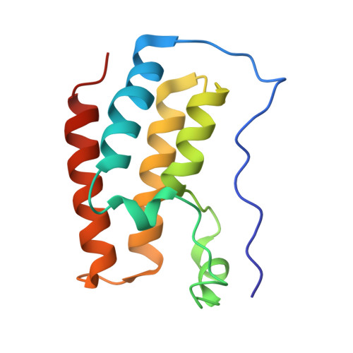
    5M39, 5M3A

  • PubMed Abstract: 

    Fragment-based drug design exploits initial screening of low molecular weight compounds and their concomitant affinity improvement. The multitude of possible chemical modifications highlights the necessity to obtain structural information about the binding mode of a fragment. Herein we describe a novel NMR methodology (LOGSY titration) that allows the determination of binding modes of low affinity binders in the protein-ligand interface and reveals suitable ligand positions for the addition of functional groups that either address or substitute protein-bound water, information of utmost importance for drug design. The particular benefit of the methodology and in contrast to conventional ligand-based methods is the independence of the molecular weight of the protein under study. The validity of the novel approach is demonstrated on two ligands interacting with bromodomain 1 of bromodomain containing protein 4, a prominent cancer target in pharmaceutical industry.


  • Organizational Affiliation

    Christian Doppler Laboratory for High-Content Structural Biology and Biotechnology, Department of Structural and Computational Biology, University of Vienna , Campus Vienna Biocenter 5, A-1030 Vienna, Austria.


Macromolecules
Find similar proteins by:  (by identity cutoff)  |  3D Structure
Entity ID: 1
MoleculeChains Sequence LengthOrganismDetailsImage
Bromodomain-containing protein 4
A, B
127Homo sapiensMutation(s): 0 
Gene Names: BRD4HUNK1
UniProt & NIH Common Fund Data Resources
Find proteins for O60885 (Homo sapiens)
Explore O60885 
Go to UniProtKB:  O60885
PHAROS:  O60885
GTEx:  ENSG00000141867 
Entity Groups  
Sequence Clusters30% Identity50% Identity70% Identity90% Identity95% Identity100% Identity
UniProt GroupO60885
Sequence Annotations
Expand
  • Reference Sequence
Small Molecules
Ligands 1 Unique
IDChains Name / Formula / InChI Key2D Diagram3D Interactions
7EA
Query on 7EA

Download Ideal Coordinates CCD File 
C [auth A],
D [auth B]
6-(3,4-dimethoxyphenyl)-3-methyl-[1,2,4]triazolo[4,3-b]pyridazine
C14 H14 N4 O2
SBNMXRKZAVLTHG-UHFFFAOYSA-N
Binding Affinity Annotations 
IDSourceBinding Affinity
7EA BindingDB:  5M39 IC50: 2.40e+4 (nM) from 1 assay(s)
Experimental Data & Validation

Experimental Data

  • Method: X-RAY DIFFRACTION
  • Resolution: 1.38 Å
  • R-Value Free: 0.242 
  • R-Value Work: 0.222 
  • R-Value Observed: 0.223 
  • Space Group: P 1
Unit Cell:
Length ( Å )Angle ( ˚ )
a = 30.379α = 99.71
b = 39.506β = 105.05
c = 57.972γ = 89.9
Software Package:
Software NamePurpose
BUSTERrefinement
XDSdata reduction
XSCALEdata scaling
PHASERphasing

Structure Validation

View Full Validation Report


Ligand Structure Quality Assessment 


Entry History 

Deposition Data

Revision History  (Full details and data files)

  • Version 1.0: 2017-09-27
    Type: Initial release
  • Version 1.1: 2017-11-22
    Changes: Database references
  • Version 1.2: 2024-01-17
    Changes: Data collection, Database references, Refinement description