


Structure of the TagL peptidoglycan binding domain from EAEC T6SS
Cambillau, C., Nguyen, V.S., Cascales, E.To be published.
Experimental Data Snapshot
wwPDB Validation 3D Report Full Report

Entity ID: 1 | |||||
|---|---|---|---|---|---|
| Molecule | Chains | Sequence Length | Organism | Details | Image |
| OmpA family protein | 139 | Escherichia coli | Mutation(s): 0 Gene Names: ACU81_04615 | 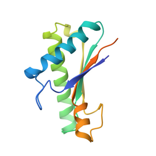 | |
UniProt | |||||
Find proteins for D3GUV9 (Escherichia coli O44:H18 (strain 042 / EAEC)) Explore D3GUV9 Go to UniProtKB: D3GUV9 | |||||
Entity Groups | |||||
| Sequence Clusters | 30% Identity50% Identity70% Identity90% Identity95% Identity100% Identity | ||||
| UniProt Group | D3GUV9 | ||||
Sequence AnnotationsExpand | |||||
| |||||
| Length ( Å ) | Angle ( ˚ ) |
|---|---|
| a = 211.55 | α = 90 |
| b = 211.55 | β = 90 |
| c = 211.55 | γ = 90 |
| Software Name | Purpose |
|---|---|
| BUSTER | refinement |
| XDS | data reduction |
| XSCALE | data scaling |
| MOLREP | phasing |
| Funding Organization | Location | Grant Number |
|---|---|---|
| French National Research Agency | France | ANR- 14-CE14-0006-01 |
RCSB PDB (citation) is hosted by
RCSB PDB is a member of the

