5M36

The molecular tweezer CLR01 stabilizes a disordered protein-protein interface


Experimental Data Snapshot

  • Method: X-RAY DIFFRACTION
  • Resolution: 2.45 Å
  • R-Value Free: 0.274 
  • R-Value Work: 0.224 
  • R-Value Observed: 0.226 

wwPDB Validation   3D Report Full Report


Ligand Structure Quality Assessment 


This is version 1.4 of the entry. See complete history


Literature

The Molecular Tweezer CLR01 Stabilizes a Disordered Protein-Protein Interface.

Bier, D.Mittal, S.Bravo-Rodriguez, K.Sowislok, A.Guillory, X.Briels, J.Heid, C.Bartel, M.Wettig, B.Brunsveld, L.Sanchez-Garcia, E.Schrader, T.Ottmann, C.

(2017) J Am Chem Soc 139: 16256-16263

  • DOI: https://doi.org/10.1021/jacs.7b07939
  • Primary Citation of Related Structures:  
    5M35, 5M36, 5M37

  • PubMed Abstract: 

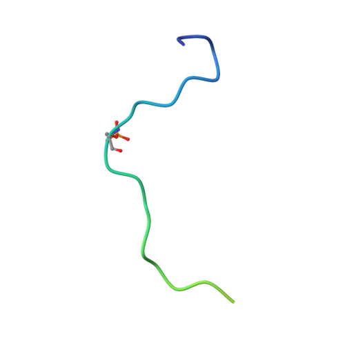
    Protein regions that are involved in protein-protein interactions (PPIs) very often display a high degree of intrinsic disorder, which is reduced during the recognition process. A prime example is binding of the rigid 14-3-3 adapter proteins to their numerous partner proteins, whose recognition motifs undergo an extensive disorder-to-order transition. In this context, it is highly desirable to control this entropy-costly process using tailored stabilizing agents. This study reveals how the molecular tweezer CLR01 tunes the 14-3-3/Cdc25CpS216 protein-protein interaction. Protein crystallography, biophysical affinity determination and biomolecular simulations unanimously deliver a remarkable finding: a supramolecular "Janus" ligand can bind simultaneously to a flexible peptidic PPI recognition motif and to a well-structured adapter protein. This binding fills a gap in the protein-protein interface, "freezes" one of the conformational states of the intrinsically disordered Cdc25C protein partner and enhances the apparent affinity of the interaction. This is the first structural and functional proof of a supramolecular ligand targeting a PPI interface and stabilizing the binding of an intrinsically disordered recognition motif to a rigid partner protein.


  • Organizational Affiliation

    Laboratory of Chemical Biology, Department of Biomedical Engineering and Institute for Complex Molecular Systems, Eindhoven University of Technology , Den Dolech 2, 5612 AZ Eindhoven, The Netherlands.


Macromolecules
Find similar proteins by:  (by identity cutoff)  |  3D Structure
Entity ID: 1
MoleculeChains Sequence LengthOrganismDetailsImage
14-3-3 protein zeta/delta
A, B
229Homo sapiensMutation(s): 0 
Gene Names: YWHAZ
UniProt & NIH Common Fund Data Resources
Find proteins for P63104 (Homo sapiens)
Explore P63104 
Go to UniProtKB:  P63104
PHAROS:  P63104
GTEx:  ENSG00000164924 
Entity Groups  
Sequence Clusters30% Identity50% Identity70% Identity90% Identity95% Identity100% Identity
UniProt GroupP63104
Sequence Annotations
Expand
  • Reference Sequence
Find similar proteins by:  (by identity cutoff)  |  3D Structure
Entity ID: 2
MoleculeChains Sequence LengthOrganismDetailsImage
M-phase inducer phosphatase 3
C, D
38Homo sapiensMutation(s): 0 
EC: 3.1.3.48
UniProt & NIH Common Fund Data Resources
Find proteins for P30307 (Homo sapiens)
Explore P30307 
Go to UniProtKB:  P30307
PHAROS:  P30307
GTEx:  ENSG00000158402 
Entity Groups  
Sequence Clusters30% Identity50% Identity70% Identity90% Identity95% Identity100% Identity
UniProt GroupP30307
Sequence Annotations
Expand
  • Reference Sequence
Small Molecules
Ligands 3 Unique
IDChains Name / Formula / InChI Key2D Diagram3D Interactions
9SZ
Query on 9SZ

Download Ideal Coordinates CCD File 
E [auth A],
F [auth A],
J [auth B],
M [auth C]
(1R,5S,9S,16R,20R,24S,28S,35R)-3,22-Bis(dihydroxyphosphoryloxy)tridecacyclo[22.14.1.15,20.19,16.128,35.02,23.04,21.06,19.08,17.010,15.025,38.027,36.029,34]dotetraconta-2(23),3,6,8(17),10,12,14,18,21,25,27(36),29,31,33,37-pentadecaene
C42 H32 O8 P2
BZESKQAEKFQSPD-FCODNRGNSA-N
BEZ
Query on BEZ

Download Ideal Coordinates CCD File 
G [auth A],
K [auth B]
BENZOIC ACID
C7 H6 O2
WPYMKLBDIGXBTP-UHFFFAOYSA-N
GOL
Query on GOL

Download Ideal Coordinates CCD File 
H [auth A],
I [auth A],
L [auth B]
GLYCEROL
C3 H8 O3
PEDCQBHIVMGVHV-UHFFFAOYSA-N
Modified Residues  2 Unique
IDChains TypeFormula2D DiagramParent
CSO
Query on CSO
A, B
L-PEPTIDE LINKINGC3 H7 N O3 SCYS
SEP
Query on SEP
C, D
L-PEPTIDE LINKINGC3 H8 N O6 PSER
Experimental Data & Validation

Experimental Data

  • Method: X-RAY DIFFRACTION
  • Resolution: 2.45 Å
  • R-Value Free: 0.274 
  • R-Value Work: 0.224 
  • R-Value Observed: 0.226 
  • Space Group: P 21 21 21
Unit Cell:
Length ( Å )Angle ( ˚ )
a = 71.52α = 90
b = 102.36β = 90
c = 112.82γ = 90
Software Package:
Software NamePurpose
REFMACrefinement
XSCALEdata scaling
PHASERphasing
PDB_EXTRACTdata extraction
XDSdata reduction
PHASERphasing

Structure Validation

View Full Validation Report


Ligand Structure Quality Assessment 


Entry History & Funding Information

Deposition Data


Funding OrganizationLocationGrant Number
German Research FoundationGermanyCollaborative Research Centre 1093
Netherlands Organisation for Scientific ResearchNetherlands024.001.035
Netherlands Organisation for Scientific ResearchNetherlands016.150.366

Revision History  (Full details and data files)

  • Version 1.0: 2017-11-15
    Type: Initial release
  • Version 1.1: 2017-11-22
    Changes: Database references
  • Version 1.2: 2018-12-12
    Changes: Data collection, Derived calculations
  • Version 1.3: 2024-01-10
    Changes: Data collection, Database references, Derived calculations, Structure summary
  • Version 1.4: 2024-01-17
    Changes: Refinement description