5M04

Structure of ObgE from Escherichia coli


Experimental Data Snapshot

  • Method: X-RAY DIFFRACTION
  • Resolution: 1.85 Å
  • R-Value Free: 0.239 
  • R-Value Work: 0.198 
  • R-Value Observed: 0.200 

wwPDB Validation   3D Report Full Report


Ligand Structure Quality Assessment 


This is version 1.3 of the entry. See complete history


Literature

Structural and biochemical analysis of Escherichia coli ObgE, a central regulator of bacterial persistence.

Gkekas, S.Singh, R.K.Shkumatov, A.V.Messens, J.Fauvart, M.Verstraeten, N.Michiels, J.Versees, W.

(2017) J Biol Chem 292: 5871-5883

  • DOI: https://doi.org/10.1074/jbc.M116.761809
  • Primary Citation of Related Structures:  
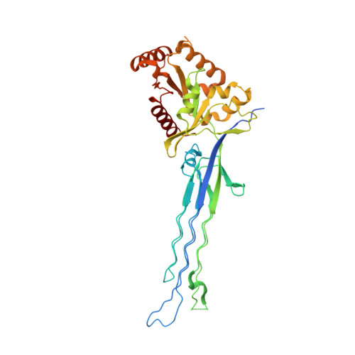
    5M04

  • PubMed Abstract: 

    The Obg protein family belongs to the TRAFAC (translation factor) class of P-loop GTPases and is conserved from bacteria to eukaryotes. Essential roles in many different cellular processes have been suggested for the Obg protein from Escherichia coli (ObgE), and we recently showed that it is a central regulator of bacterial persistence. Here, we report the first crystal structure of ObgE at 1.85-Å resolution in the GDP-bound state, showing the characteristic N-terminal domain and a central G domain that are common to all Obg proteins. ObgE also contains an intrinsically disordered C-terminal domain, and we show here that this domain specifically contributed to GTP binding, whereas it did not influence GDP binding or GTP hydrolysis. Biophysical analysis, using small angle X-ray scattering and multi-angle light scattering experiments, revealed that ObgE is a monomer in solution, regardless of the bound nucleotide. In contrast to recent suggestions, our biochemical analyses further indicate that ObgE is neither activated by K + ions nor by homodimerization. However, the ObgE GTPase activity was stimulated upon binding to the ribosome, confirming the ribosome-dependent GTPase activity of the Obg family. Combined, our data represent an important step toward further unraveling the detailed molecular mechanism of ObgE, which might pave the way to further studies into how this GTPase regulates bacterial physiology, including persistence.


  • Organizational Affiliation

    From the Structural Biology Brussels, Vrije Universiteit Brussel, 1050 Brussels.


Macromolecules
Find similar proteins by:  (by identity cutoff)  |  3D Structure
Entity ID: 1
MoleculeChains Sequence LengthOrganismDetailsImage
GTPase ObgE/CgtA360Escherichia coli DH5[alpha]Mutation(s): 0 
Gene Names: obgEcgtAobgyhbZb3183JW3150
EC: 3.6.5
UniProt
Find proteins for P42641 (Escherichia coli (strain K12))
Explore P42641 
Go to UniProtKB:  P42641
Entity Groups  
Sequence Clusters30% Identity50% Identity70% Identity90% Identity95% Identity100% Identity
UniProt GroupP42641
Sequence Annotations
Expand
  • Reference Sequence
Small Molecules
Ligands 2 Unique
IDChains Name / Formula / InChI Key2D Diagram3D Interactions
GDP
Query on GDP

Download Ideal Coordinates CCD File 
B [auth A]GUANOSINE-5'-DIPHOSPHATE
C10 H15 N5 O11 P2
QGWNDRXFNXRZMB-UUOKFMHZSA-N
MG
Query on MG

Download Ideal Coordinates CCD File 
C [auth A]MAGNESIUM ION
Mg
JLVVSXFLKOJNIY-UHFFFAOYSA-N
Binding Affinity Annotations 
IDSourceBinding Affinity
GDP Binding MOAD:  5M04 Kd: 460 (nM) from 1 assay(s)
Experimental Data & Validation

Experimental Data

  • Method: X-RAY DIFFRACTION
  • Resolution: 1.85 Å
  • R-Value Free: 0.239 
  • R-Value Work: 0.198 
  • R-Value Observed: 0.200 
  • Space Group: C 2 2 21
Unit Cell:
Length ( Å )Angle ( ˚ )
a = 64.59α = 90
b = 83β = 90
c = 177.46γ = 90
Software Package:
Software NamePurpose
REFMACrefinement
XDSdata reduction
XDSdata scaling
PHASERphasing

Structure Validation

View Full Validation Report


Ligand Structure Quality Assessment 


Entry History & Funding Information

Deposition Data


Funding OrganizationLocationGrant Number
Fonds voor Wetenschappelijk Onderzoek (FWO)BelgiumG.0471.12N, G0B2515N
Institute for the Promotion of Innovation through Science and Technology in Flanders (IWT).Belgium--

Revision History  (Full details and data files)

  • Version 1.0: 2017-03-01
    Type: Initial release
  • Version 1.1: 2017-03-08
    Changes: Database references
  • Version 1.2: 2017-04-19
    Changes: Database references
  • Version 1.3: 2024-01-17
    Changes: Data collection, Database references, Refinement description