5LZ3

Crystal structure of human ACBD3 GOLD domain in complex with 3A protein of Aichivirus A


Experimental Data Snapshot

  • Method: X-RAY DIFFRACTION
  • Resolution: 3.00 Å
  • R-Value Free: 0.248 
  • R-Value Work: 0.218 
  • R-Value Observed: 0.220 

wwPDB Validation   3D Report Full Report


This is version 1.2 of the entry. See complete history


Literature

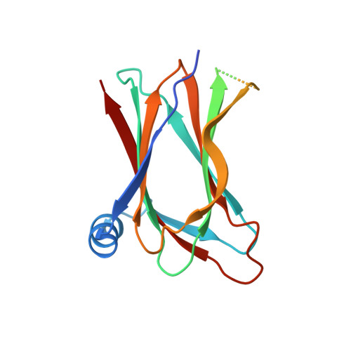
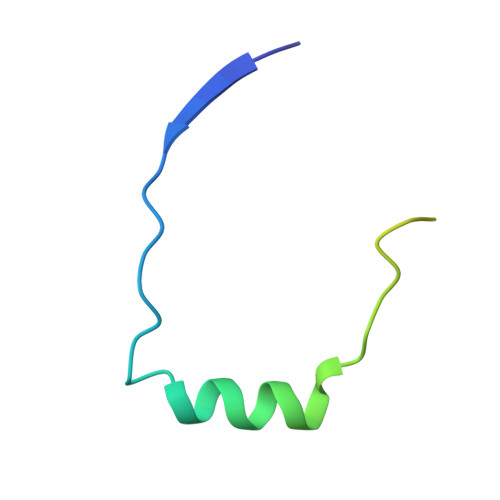
Kobuviral Non-structural 3A Proteins Act as Molecular Harnesses to Hijack the Host ACBD3 Protein.

Klima, M.Chalupska, D.Rozycki, B.Humpolickova, J.Rezabkova, L.Silhan, J.Baumlova, A.Dubankova, A.Boura, E.

(2017) Structure 25: 219-230

  • DOI: https://doi.org/10.1016/j.str.2016.11.021
  • Primary Citation of Related Structures:  
    5LZ1, 5LZ3, 5LZ6

  • PubMed Abstract: 

    Picornaviruses are small positive-sense single-stranded RNA viruses that include many important human pathogens. Within the host cell, they replicate at specific replication sites called replication organelles. To create this membrane platform, they hijack several host factors including the acyl-CoA-binding domain-containing protein-3 (ACBD3). Here, we present a structural characterization of the molecular complexes formed by the non-structural 3A proteins from two species of the Kobuvirus genus of the Picornaviridae family and the 3A-binding domain of the host ACBD3 protein. Specifically, we present a series of crystal structures as well as a molecular dynamics simulation of the 3A:ACBD3 complex at the membrane, which reveals that the viral 3A proteins act as molecular harnesses to enslave the ACBD3 protein leading to its stabilization at target membranes. Our data provide a structural rationale for understanding how these viral-host protein complexes assemble at the atomic level and identify new potential targets for antiviral therapies.


  • Organizational Affiliation

    Institute of Organic Chemistry and Biochemistry, Czech Academy of Sciences, 16610 Prague, Czech Republic. Electronic address: klima@uochb.cas.cz.


Macromolecules
Find similar proteins by:  (by identity cutoff)  |  3D Structure
Entity ID: 1
MoleculeChains Sequence LengthOrganismDetailsImage
Golgi resident protein GCP60166Homo sapiensMutation(s): 0 
Gene Names: ACBD3GCP60GOCAP1GOLPH1
UniProt & NIH Common Fund Data Resources
Find proteins for Q9H3P7 (Homo sapiens)
Explore Q9H3P7 
Go to UniProtKB:  Q9H3P7
PHAROS:  Q9H3P7
GTEx:  ENSG00000182827 
Entity Groups  
Sequence Clusters30% Identity50% Identity70% Identity90% Identity95% Identity100% Identity
UniProt GroupQ9H3P7
Sequence Annotations
Expand
  • Reference Sequence
Find similar proteins by:  (by identity cutoff)  |  3D Structure
Entity ID: 2
MoleculeChains Sequence LengthOrganismDetailsImage
3A58Aichivirus AMutation(s): 0 
UniProt
Find proteins for O91464 (Aichi virus (strain Human/A846/88/1989))
Explore O91464 
Go to UniProtKB:  O91464
Entity Groups  
Sequence Clusters30% Identity50% Identity70% Identity90% Identity95% Identity100% Identity
UniProt GroupO91464
Sequence Annotations
Expand
  • Reference Sequence
Experimental Data & Validation

Experimental Data

  • Method: X-RAY DIFFRACTION
  • Resolution: 3.00 Å
  • R-Value Free: 0.248 
  • R-Value Work: 0.218 
  • R-Value Observed: 0.220 
  • Space Group: I 2 2 2
Unit Cell:
Length ( Å )Angle ( ˚ )
a = 54.1α = 90
b = 81.15β = 90
c = 117.57γ = 90
Software Package:
Software NamePurpose
PHENIXrefinement
XDSdata reduction
XDSdata scaling
PHASERphasing

Structure Validation

View Full Validation Report


Entry History 

Deposition Data

Revision History  (Full details and data files)

  • Version 1.0: 2016-12-14
    Type: Initial release
  • Version 1.1: 2017-12-13
    Changes: Database references
  • Version 1.2: 2024-01-17
    Changes: Data collection, Database references, Refinement description