5LXM

Crystal structure of Aurora-A bound to a hydrocarbon-stapled proteomimetic of TPX2


Experimental Data Snapshot

  • Method: X-RAY DIFFRACTION
  • Resolution: 2.08 Å
  • R-Value Free: 0.243 
  • R-Value Work: 0.200 
  • R-Value Observed: 0.202 

wwPDB Validation   3D Report Full Report


Ligand Structure Quality Assessment 


This is version 1.4 of the entry. See complete history


Literature

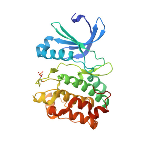
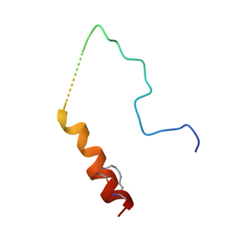
A TPX2 Proteomimetic Has Enhanced Affinity for Aurora-A Due to Hydrocarbon Stapling of a Helix.

Rennie, Y.K.McIntyre, P.J.Akindele, T.Bayliss, R.Jamieson, A.G.

(2016) ACS Chem Biol 11: 3383-3390

  • DOI: https://doi.org/10.1021/acschembio.6b00727
  • Primary Citation of Related Structures:  
    5LXM

  • PubMed Abstract: 

    Inhibition of protein kinases using ATP-competitive compounds is an important strategy in drug discovery. In contrast, the allosteric regulation of kinases through the disruption of protein-protein interactions has not been widely adopted, despite the potential for selective targeting. Aurora-A kinase regulates mitotic entry and mitotic spindle assembly and is a promising target for anticancer therapy. The microtubule-associated protein TPX2 activates Aurora-A through binding to two sites. Aurora-A recognition is mediated by two motifs within the first 43 residues of TPX2, connected by a flexible linker. To characterize the contributions of these three structural elements, we prepared a series of TPX2 proteomimetics and investigated their binding affinity for Aurora-A using isothermal titration calorimetry. A novel stapled TPX2 peptide was developed that has improved binding affinity for Aurora-A and mimics the function of TPX2 in activating Aurora-A's autophosphorylation. We conclude that the helical region of TPX2 folds upon binding Aurora-A, and that stabilization of this helix does not compromise Aurora-A activation. This study demonstrates that the preparation of these proteomimetics using modern synthesis methods is feasible and their biochemical evaluation demonstrates the power of proteomimetics as tool compounds for investigating PPIs involving intrinsically disordered regions of proteins.


  • Organizational Affiliation

    Department of Chemistry, University of Leicester , Lancaster Road, Leicester LE1 9HN, United Kingdom.


Macromolecules
Find similar proteins by:  (by identity cutoff)  |  3D Structure
Entity ID: 1
MoleculeChains Sequence LengthOrganismDetailsImage
Aurora kinase A283Homo sapiensMutation(s): 2 
Gene Names: AURKAAIKAIRK1ARK1AURAAYK1BTAKIAK1STK15STK6
EC: 2.7.11.1
UniProt & NIH Common Fund Data Resources
Find proteins for O14965 (Homo sapiens)
Explore O14965 
Go to UniProtKB:  O14965
PHAROS:  O14965
GTEx:  ENSG00000087586 
Entity Groups  
Sequence Clusters30% Identity50% Identity70% Identity90% Identity95% Identity100% Identity
UniProt GroupO14965
Sequence Annotations
Expand
  • Reference Sequence
Find similar proteins by:  (by identity cutoff)  |  3D Structure
Entity ID: 2
MoleculeChains Sequence LengthOrganismDetailsImage
Targeting protein for Xklp2B [auth D]38Homo sapiensMutation(s): 0 
UniProt & NIH Common Fund Data Resources
Find proteins for Q9ULW0 (Homo sapiens)
Explore Q9ULW0 
Go to UniProtKB:  Q9ULW0
PHAROS:  Q9ULW0
GTEx:  ENSG00000088325 
Entity Groups  
Sequence Clusters30% Identity50% Identity70% Identity90% Identity95% Identity100% Identity
UniProt GroupQ9ULW0
Sequence Annotations
Expand
  • Reference Sequence
Small Molecules
Ligands 9 Unique
IDChains Name / Formula / InChI Key2D Diagram3D Interactions
ADP
Query on ADP

Download Ideal Coordinates CCD File 
C [auth A]ADENOSINE-5'-DIPHOSPHATE
C10 H15 N5 O10 P2
XTWYTFMLZFPYCI-KQYNXXCUSA-N
MES
Query on MES

Download Ideal Coordinates CCD File 
H [auth A]2-(N-MORPHOLINO)-ETHANESULFONIC ACID
C6 H13 N O4 S
SXGZJKUKBWWHRA-UHFFFAOYSA-N
PG4
Query on PG4

Download Ideal Coordinates CCD File 
D [auth A]TETRAETHYLENE GLYCOL
C8 H18 O5
UWHCKJMYHZGTIT-UHFFFAOYSA-N
P4G
Query on P4G

Download Ideal Coordinates CCD File 
I [auth A]1-ETHOXY-2-(2-ETHOXYETHOXY)ETHANE
C8 H18 O3
RRQYJINTUHWNHW-UHFFFAOYSA-N
PEG
Query on PEG

Download Ideal Coordinates CCD File 
F [auth A],
G [auth A]
DI(HYDROXYETHYL)ETHER
C4 H10 O3
MTHSVFCYNBDYFN-UHFFFAOYSA-N
SO4
Query on SO4

Download Ideal Coordinates CCD File 
E [auth A]SULFATE ION
O4 S
QAOWNCQODCNURD-UHFFFAOYSA-L
CL
Query on CL

Download Ideal Coordinates CCD File 
J [auth A]CHLORIDE ION
Cl
VEXZGXHMUGYJMC-UHFFFAOYSA-M
MG
Query on MG

Download Ideal Coordinates CCD File 
K [auth A],
L [auth A]
MAGNESIUM ION
Mg
JLVVSXFLKOJNIY-UHFFFAOYSA-N
NH3
Query on NH3

Download Ideal Coordinates CCD File 
M [auth D]AMMONIA
H3 N
QGZKDVFQNNGYKY-UHFFFAOYSA-N
Modified Residues  2 Unique
IDChains TypeFormula2D DiagramParent
TPO
Query on TPO
A
L-PEPTIDE LINKINGC4 H10 N O6 PTHR
MK8
Query on MK8
B [auth D]L-PEPTIDE LINKINGC7 H15 N O2LEU
Experimental Data & Validation

Experimental Data

  • Method: X-RAY DIFFRACTION
  • Resolution: 2.08 Å
  • R-Value Free: 0.243 
  • R-Value Work: 0.200 
  • R-Value Observed: 0.202 
  • Space Group: P 31 2 1
Unit Cell:
Length ( Å )Angle ( ˚ )
a = 79.78α = 90
b = 79.78β = 90
c = 140.26γ = 120
Software Package:
Software NamePurpose
PHENIXrefinement
PDB_EXTRACTdata extraction
xia2data reduction
xia2data scaling

Structure Validation

View Full Validation Report


Ligand Structure Quality Assessment 


Entry History & Funding Information

Deposition Data


Funding OrganizationLocationGrant Number
Medical Research Council (United Kingdom)United KingdomMR/K016903/1
Cancer Research UKUnited KingdomC24461/A12772

Revision History  (Full details and data files)

  • Version 1.0: 2016-11-02
    Type: Initial release
  • Version 1.1: 2016-12-28
    Changes: Database references
  • Version 1.2: 2017-09-13
    Changes: Advisory, Author supporting evidence
  • Version 1.3: 2019-10-16
    Changes: Data collection
  • Version 1.4: 2024-02-07
    Changes: Advisory, Data collection, Database references, Derived calculations, Refinement description