5LXC

Crystal structure of DYRK2 in complex with EHT 5372 (Compound 1)


Experimental Data Snapshot

  • Method: X-RAY DIFFRACTION
  • Resolution: 2.15 Å
  • R-Value Free: 0.254 
  • R-Value Work: 0.206 
  • R-Value Observed: 0.208 

wwPDB Validation   3D Report Full Report


Ligand Structure Quality Assessment 


This is version 1.1 of the entry. See complete history


Literature

An Unusual Binding Model of the Methyl 9-Anilinothiazolo[5,4-f] quinazoline-2-carbimidates (EHT 1610 and EHT 5372) Confers High Selectivity for Dual-Specificity Tyrosine Phosphorylation-Regulated Kinases.

Chaikuad, A.Diharce, J.Schroder, M.Foucourt, A.Leblond, B.Casagrande, A.S.Desire, L.Bonnet, P.Knapp, S.Besson, T.

(2016) J Med Chem 59: 10315-10321

  • DOI: https://doi.org/10.1021/acs.jmedchem.6b01083
  • Primary Citation of Related Structures:  
    5LXC, 5LXD

  • PubMed Abstract: 

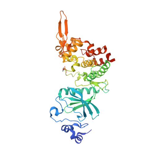
    Methyl 9-anilinothiazolo[5,4-f]quinazoline-2-carbimidates 1 (EHT 5372) and 2 (EHT 1610) are strong inhibitors of DYRK's family kinases. The crystal structures of the complex revealed a noncanonical binding mode of compounds 1 and 2 in DYRK2, explaining the remarkable selectivity and potency of these inhibitors. The structural data and comparison presented here provide therefore a template for further improvement of this inhibitor class and for the development of novel inhibitors selectively targeting DYRK kinases.


  • Organizational Affiliation

    Target Discovery Institute (TDI), and Structural Genomics Consortium (SGC), University of Oxford , Old Road Campus Research Building, Oxford OX3 7DQ, U.K.


Macromolecules
Find similar proteins by:  (by identity cutoff)  |  3D Structure
Entity ID: 1
MoleculeChains Sequence LengthOrganismDetailsImage
Dual specificity tyrosine-phosphorylation-regulated kinase 2
A, B
408Homo sapiensMutation(s): 0 
Gene Names: DYRK2
EC: 2.7.12.1
UniProt & NIH Common Fund Data Resources
Find proteins for Q92630 (Homo sapiens)
Explore Q92630 
Go to UniProtKB:  Q92630
PHAROS:  Q92630
GTEx:  ENSG00000127334 
Entity Groups  
Sequence Clusters30% Identity50% Identity70% Identity90% Identity95% Identity100% Identity
UniProt GroupQ92630
Sequence Annotations
Expand
  • Reference Sequence
Small Molecules
Ligands 2 Unique
IDChains Name / Formula / InChI Key2D Diagram3D Interactions
7AA
Query on 7AA

Download Ideal Coordinates CCD File 
C [auth A],
L [auth B]
methyl 9-[(2,4-dichlorophenyl)amino]-[1,3]thiazolo[5,4-f]quinazoline-2-carboximidate
C17 H11 Cl2 N5 O S
QSGKPYRFWJINEH-HMMYKYKNSA-N
EDO
Query on EDO

Download Ideal Coordinates CCD File 
D [auth A]
E [auth A]
F [auth A]
G [auth A]
H [auth A]
D [auth A],
E [auth A],
F [auth A],
G [auth A],
H [auth A],
I [auth A],
J [auth A],
K [auth A],
M [auth B],
N [auth B],
O [auth B]
1,2-ETHANEDIOL
C2 H6 O2
LYCAIKOWRPUZTN-UHFFFAOYSA-N
Modified Residues  1 Unique
IDChains TypeFormula2D DiagramParent
PTR
Query on PTR
A, B
L-PEPTIDE LINKINGC9 H12 N O6 PTYR
Binding Affinity Annotations 
IDSourceBinding Affinity
7AA Binding MOAD:  5LXC IC50: 10.8 (nM) from 1 assay(s)
Experimental Data & Validation

Experimental Data

  • Method: X-RAY DIFFRACTION
  • Resolution: 2.15 Å
  • R-Value Free: 0.254 
  • R-Value Work: 0.206 
  • R-Value Observed: 0.208 
  • Space Group: C 1 2 1
Unit Cell:
Length ( Å )Angle ( ˚ )
a = 130.22α = 90
b = 60.98β = 105.04
c = 148.79γ = 90
Software Package:
Software NamePurpose
REFMACrefinement
iMOSFLMdata reduction
SCALAdata scaling
PHASERphasing

Structure Validation

View Full Validation Report


Ligand Structure Quality Assessment 


Entry History 

Deposition Data

Revision History  (Full details and data files)

  • Version 1.0: 2016-10-26
    Type: Initial release
  • Version 1.1: 2017-01-11
    Changes: Database references