5LWN

Crystal structure of JAK3 in complex with Compound 5 (FM409)


Experimental Data Snapshot

  • Method: X-RAY DIFFRACTION
  • Resolution: 1.60 Å
  • R-Value Free: 0.209 
  • R-Value Work: 0.166 
  • R-Value Observed: 0.169 

wwPDB Validation   3D Report Full Report


Ligand Structure Quality Assessment 


This is version 1.1 of the entry. See complete history


Literature

Selective JAK3 Inhibitors with a Covalent Reversible Binding Mode Targeting a New Induced Fit Binding Pocket.

Forster, M.Chaikuad, A.Bauer, S.M.Holstein, J.Robers, M.B.Corona, C.R.Gehringer, M.Pfaffenrot, E.Ghoreschi, K.Knapp, S.Laufer, S.A.

(2016) Cell Chem Biol 23: 1335-1340

  • DOI: https://doi.org/10.1016/j.chembiol.2016.10.008
  • Primary Citation of Related Structures:  
    5LWM, 5LWN

  • PubMed Abstract: 

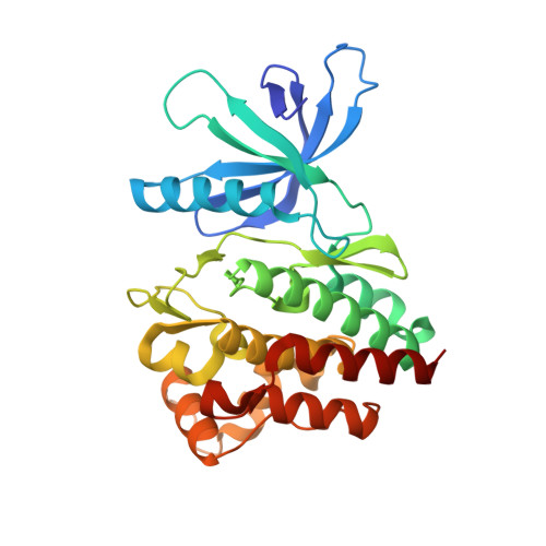
    Janus kinases (JAKs) are a family of cytoplasmatic tyrosine kinases that are attractive targets for the development of anti-inflammatory drugs given their roles in cytokine signaling. One question regarding JAKs and their inhibitors that remains under intensive debate is whether JAK inhibitors should be isoform selective. Since JAK3 functions are restricted to immune cells, an isoform-selective inhibitor for JAK3 could be especially valuable to achieve clinically more useful and precise effects. However, the high degree of structural conservation makes isoform-selective targeting a challenging task. Here, we present picomolar inhibitors with unprecedented kinome-wide selectivity for JAK3. Selectivity was achieved by concurrent covalent reversible targeting of a JAK3-specific cysteine residue and a ligand-induced binding pocket. We confirmed that in vitro activity and selectivity translate well into the cellular environment and suggest that our inhibitors are powerful tools to elucidate JAK3-specific functions.


  • Organizational Affiliation

    Department of Pharmaceutical/Medicinal Chemistry, Eberhard-Karls-University Tuebingen, Auf der Morgenstelle 8, 72076 Tuebingen, Germany.


Macromolecules
Find similar proteins by:  (by identity cutoff)  |  3D Structure
Entity ID: 1
MoleculeChains Sequence LengthOrganismDetailsImage
Tyrosine-protein kinase JAK3294Homo sapiensMutation(s): 0 
Gene Names: JAK3
EC: 2.7.10.2
UniProt & NIH Common Fund Data Resources
Find proteins for P52333 (Homo sapiens)
Explore P52333 
Go to UniProtKB:  P52333
PHAROS:  P52333
GTEx:  ENSG00000105639 
Entity Groups  
Sequence Clusters30% Identity50% Identity70% Identity90% Identity95% Identity100% Identity
UniProt GroupP52333
Sequence Annotations
Expand
  • Reference Sequence
Small Molecules
Ligands 4 Unique
IDChains Name / Formula / InChI Key2D Diagram3D Interactions
79S
Query on 79S

Download Ideal Coordinates CCD File 
J [auth A](2~{S})-2-cyano-~{N},~{N}-dimethyl-3-[5-[3-[(1~{S},2~{R})-2-methylcyclohexyl]-3,5,8,10-tetrazatricyclo[7.3.0.0^{2,6}]dodeca-1,4,6,8,11-pentaen-4-yl]furan-2-yl]propanamide
C25 H28 N6 O2
AMJNKQADUVRGCR-GUXCAODWSA-N
79R
Query on 79R

Download Ideal Coordinates CCD File 
I [auth A](~{Z})-2-cyano-~{N},~{N}-dimethyl-3-[5-[3-[(1~{S},2~{R})-2-methylcyclohexyl]-3,5,8,10-tetrazatricyclo[7.3.0.0^{2,6}]dodeca-1,4,6,8,11-pentaen-4-yl]furan-2-yl]prop-2-enamide
C25 H26 N6 O2
LVVYJCDRPPCFEQ-YXEVWGHGSA-N
PHU
Query on PHU

Download Ideal Coordinates CCD File 
B [auth A]1-phenylurea
C7 H8 N2 O
LUBJCRLGQSPQNN-UHFFFAOYSA-N
EDO
Query on EDO

Download Ideal Coordinates CCD File 
C [auth A]
D [auth A]
E [auth A]
F [auth A]
G [auth A]
C [auth A],
D [auth A],
E [auth A],
F [auth A],
G [auth A],
H [auth A]
1,2-ETHANEDIOL
C2 H6 O2
LYCAIKOWRPUZTN-UHFFFAOYSA-N
Binding Affinity Annotations 
IDSourceBinding Affinity
79R Binding MOAD:  5LWN IC50: 17 (nM) from 1 assay(s)
Experimental Data & Validation

Experimental Data

  • Method: X-RAY DIFFRACTION
  • Resolution: 1.60 Å
  • R-Value Free: 0.209 
  • R-Value Work: 0.166 
  • R-Value Observed: 0.169 
  • Space Group: P 1 21 1
Unit Cell:
Length ( Å )Angle ( ˚ )
a = 42.049α = 90
b = 62.57β = 92.55
c = 51.08γ = 90
Software Package:
Software NamePurpose
REFMACrefinement
iMOSFLMdata reduction
SCALAdata scaling
PHASERphasing

Structure Validation

View Full Validation Report


Ligand Structure Quality Assessment 


Entry History 

Revision History  (Full details and data files)

  • Version 1.0: 2016-10-26
    Type: Initial release
  • Version 1.1: 2016-11-30
    Changes: Database references