5LRT

Structure of the Deamidase-Depupylase Dop of the Prokaryotic Ubiquitin-like Modification Pathway in Complex with ADP and Phosphate


Experimental Data Snapshot

  • Method: X-RAY DIFFRACTION
  • Resolution: 1.85 Å
  • R-Value Free: 0.198 
  • R-Value Work: 0.167 
  • R-Value Observed: 0.168 

wwPDB Validation   3D Report Full Report


Ligand Structure Quality Assessment 


This is version 1.3 of the entry. See complete history


Literature

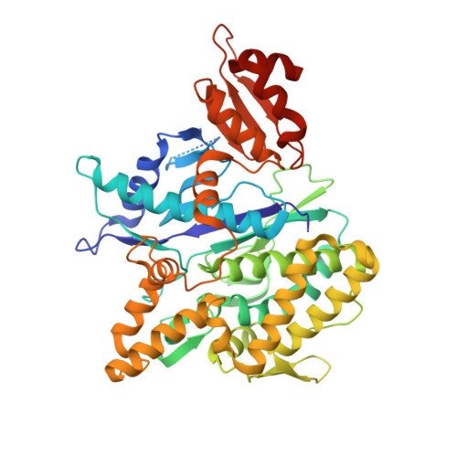
Depupylase Dop Requires Inorganic Phosphate in the Active Site for Catalysis.

Bolten, M.Vahlensieck, C.Lipp, C.Leibundgut, M.Ban, N.Weber-Ban, E.

(2017) J Biol Chem 292: 4044-4053

  • DOI: https://doi.org/10.1074/jbc.M116.755645
  • Primary Citation of Related Structures:  
    5LRT

  • PubMed Abstract: 

    Analogous to eukaryotic ubiquitination, proteins in actinobacteria can be post-translationally modified in a process referred to as pupylation, the covalent attachment of prokaryotic ubiquitin-like protein Pup to lysine side chains of the target protein via an isopeptide bond. As in eukaryotes, an opposing activity counteracts the modification by specific cleavage of the isopeptide bond formed with Pup. However, the enzymes involved in pupylation and depupylation have evolved independently of ubiquitination and are related to the family of ATP-binding and hydrolyzing carboxylate-amine ligases of the glutamine synthetase type. Furthermore, the Pup ligase PafA and the depupylase Dop share close structural and sequence homology and have a common evolutionary history despite catalyzing opposing reactions. Here, we investigate the role played by the nucleotide in the active site of the depupylase Dop using a combination of biochemical experiments and X-ray crystallographic studies. We show that, although Dop does not turn over ATP stoichiometrically with substrate, the active site nucleotide species in Dop is ADP and inorganic phosphate rather than ATP, and that non-hydrolyzable analogs of ATP cannot support the enzymatic reaction. This finding suggests that the catalytic mechanism is more similar to the mechanism of the ligase PafA than previously thought and likely involves the transient formation of a phosphorylated Pup-intermediate. Evidence is presented for a mechanism where the inorganic phosphate acts as the nucleophilic species in amide bond cleavage and implications for Dop function are discussed.


  • Organizational Affiliation

    From the ETH Zurich, Institute of Molecular Biology & Biophysics, 8093 Zurich, Switzerland.


Macromolecules
Find similar proteins by:  (by identity cutoff)  |  3D Structure
Entity ID: 1
MoleculeChains Sequence LengthOrganismDetailsImage
Depupylase508Acidothermus cellulolyticus 11BMutation(s): 0 
Gene Names: dopAcel_1186
EC: 3.4
UniProt
Find proteins for A0LU48 (Acidothermus cellulolyticus (strain ATCC 43068 / DSM 8971 / 11B))
Explore A0LU48 
Go to UniProtKB:  A0LU48
Entity Groups  
Sequence Clusters30% Identity50% Identity70% Identity90% Identity95% Identity100% Identity
UniProt GroupA0LU48
Sequence Annotations
Expand
  • Reference Sequence
Small Molecules
Ligands 7 Unique
IDChains Name / Formula / InChI Key2D Diagram3D Interactions
ADP
Query on ADP

Download Ideal Coordinates CCD File 
B [auth A]ADENOSINE-5'-DIPHOSPHATE
C10 H15 N5 O10 P2
XTWYTFMLZFPYCI-KQYNXXCUSA-N
P6G
Query on P6G

Download Ideal Coordinates CCD File 
H [auth A]HEXAETHYLENE GLYCOL
C12 H26 O7
IIRDTKBZINWQAW-UHFFFAOYSA-N
PGE
Query on PGE

Download Ideal Coordinates CCD File 
I [auth A],
J [auth A]
TRIETHYLENE GLYCOL
C6 H14 O4
ZIBGPFATKBEMQZ-UHFFFAOYSA-N
PEG
Query on PEG

Download Ideal Coordinates CCD File 
K [auth A]DI(HYDROXYETHYL)ETHER
C4 H10 O3
MTHSVFCYNBDYFN-UHFFFAOYSA-N
PO4
Query on PO4

Download Ideal Coordinates CCD File 
G [auth A]PHOSPHATE ION
O4 P
NBIIXXVUZAFLBC-UHFFFAOYSA-K
MG
Query on MG

Download Ideal Coordinates CCD File 
C [auth A],
D [auth A],
E [auth A],
F [auth A]
MAGNESIUM ION
Mg
JLVVSXFLKOJNIY-UHFFFAOYSA-N
NA
Query on NA

Download Ideal Coordinates CCD File 
L [auth A]SODIUM ION
Na
FKNQFGJONOIPTF-UHFFFAOYSA-N
Experimental Data & Validation

Experimental Data

  • Method: X-RAY DIFFRACTION
  • Resolution: 1.85 Å
  • R-Value Free: 0.198 
  • R-Value Work: 0.167 
  • R-Value Observed: 0.168 
  • Space Group: P 31 2 1
Unit Cell:
Length ( Å )Angle ( ˚ )
a = 72.398α = 90
b = 72.398β = 90
c = 215.25γ = 120
Software Package:
Software NamePurpose
Aimlessdata scaling
PHASERphasing
PHENIXrefinement
PDB_EXTRACTdata extraction
XDSdata reduction

Structure Validation

View Full Validation Report


Ligand Structure Quality Assessment 


Entry History 

Deposition Data

Revision History  (Full details and data files)

  • Version 1.0: 2017-02-01
    Type: Initial release
  • Version 1.1: 2017-02-08
    Changes: Database references
  • Version 1.2: 2017-03-22
    Changes: Database references
  • Version 1.3: 2024-01-17
    Changes: Data collection, Database references, Derived calculations, Refinement description