


Crystal structure of the de-sumoylating protease
Eckhoff, J., Dohmen, J., Pichlo, C., Baumann, U.To be published.
Experimental Data Snapshot
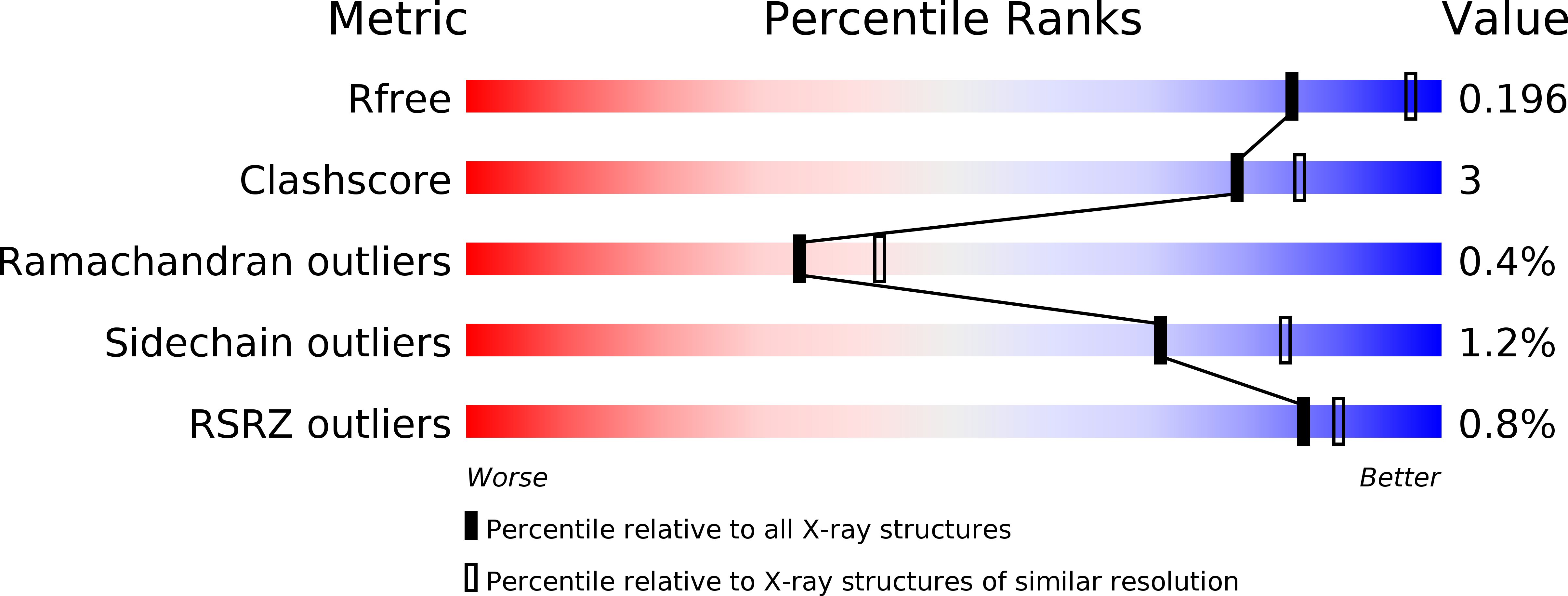
wwPDB Validation 3D Report Full Report
Entity ID: 1 | |||||
|---|---|---|---|---|---|
| Molecule | Chains | Sequence Length | Organism | Details | Image |
| Ubiquitin-like-specific protease 2 | A [auth B] | 310 | Saccharomyces cerevisiae S288C | Mutation(s): 0 Gene Names: ULP2, SMT4, YIL031W EC: 3.4.22.68 | 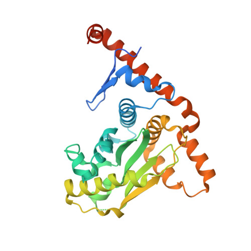 |
UniProt | |||||
Find proteins for P40537 (Saccharomyces cerevisiae (strain ATCC 204508 / S288c)) Explore P40537 Go to UniProtKB: P40537 | |||||
Entity Groups | |||||
| Sequence Clusters | 30% Identity50% Identity70% Identity90% Identity95% Identity100% Identity | ||||
| UniProt Group | P40537 | ||||
Sequence AnnotationsExpand | |||||
| |||||
| Ligands 1 Unique | |||||
|---|---|---|---|---|---|
| ID | Chains | Name / Formula / InChI Key | 2D Diagram | 3D Interactions | |
| ACT Query on ACT | B | ACETATE ION C2 H3 O2 QTBSBXVTEAMEQO-UHFFFAOYSA-M |  | ||
| Length ( Å ) | Angle ( ˚ ) |
|---|---|
| a = 42.54 | α = 90 |
| b = 55.58 | β = 90 |
| c = 172.78 | γ = 90 |
| Software Name | Purpose |
|---|---|
| PHENIX | refinement |
| XDS | data reduction |
| XSCALE | data scaling |
| PHENIX | phasing |
| Funding Organization | Location | Grant Number |
|---|---|---|
| German Research Foundation | Germany | -- |
RCSB PDB (citation) is hosted by
RCSB PDB is a member of the

