Structure and genome release of Twort-like Myoviridae phage with a double-layered baseplate.
Novacek, J., Siborova, M., Benesik, M., Pantucek, R., Doskar, J., Plevka, P.(2016) Proc Natl Acad Sci U S A 113: 9351-9356
- PubMed: 27469164
- DOI: https://doi.org/10.1073/pnas.1605883113
- Primary Citation of Related Structures:
5LI2, 5LI4, 5LII, 5LIJ - PubMed Abstract:
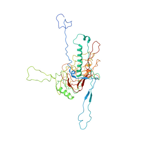
Bacteriophages from the family Myoviridae use double-layered contractile tails to infect bacteria. Contraction of the tail sheath enables the tail tube to penetrate through the bacterial cell wall and serve as a channel for the transport of the phage genome into the cytoplasm. However, the mechanisms controlling the tail contraction and genome release of phages with "double-layered" baseplates were unknown. We used cryo-electron microscopy to show that the binding of the Twort-like phage phi812 to the Staphylococcus aureus cell wall requires a 210° rotation of the heterohexameric receptor-binding and tripod protein complexes within its baseplate about an axis perpendicular to the sixfold axis of the tail. This rotation reorients the receptor-binding proteins to point away from the phage head, and also results in disruption of the interaction of the tripod proteins with the tail sheath, hence triggering its contraction. However, the tail sheath contraction of Myoviridae phages is not sufficient to induce genome ejection. We show that the end of the phi812 double-stranded DNA genome is bound to one protein subunit from a connector complex that also forms an interface between the phage head and tail. The tail sheath contraction induces conformational changes of the neck and connector that result in disruption of the DNA binding. The genome penetrates into the neck, but is stopped at a bottleneck before the tail tube. A subsequent structural change of the tail tube induced by its interaction with the S. aureus cell is required for the genome's release.
Organizational Affiliation:
Central European Institute of Technology, Masaryk University, 625 00 Brno, Czech Republic;