5LGC

T48 deacetylase with substrate


Experimental Data Snapshot

  • Method: X-RAY DIFFRACTION
  • Resolution: 2.09 Å
  • R-Value Free: 0.223 
  • R-Value Work: 0.185 
  • R-Value Observed: 0.187 

wwPDB Validation   3D Report Full Report


This is version 2.1 of the entry. See complete history


Literature

Structure and function of a CE4 deacetylase isolated from a marine environment.

Tuveng, T.R.Rothweiler, U.Udatha, G.Vaaje-Kolstad, G.Smalas, A.Eijsink, V.G.H.

(2017) PLoS One 12: e0187544-e0187544

  • DOI: https://doi.org/10.1371/journal.pone.0187544
  • Primary Citation of Related Structures:  
    5LFZ, 5LGC

  • PubMed Abstract: 

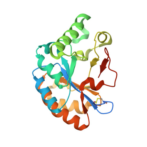
    Chitin, a polymer of β(1-4)-linked N-acetylglucosamine found in e.g. arthropods, is a valuable resource that may be used to produce chitosan and chitooligosaccharides, two compounds with considerable industrial and biomedical potential. Deacetylating enzymes may be used to tailor the properties of chitin and its derived products. Here, we describe a novel CE4 enzyme originating from a marine Arthrobacter species (ArCE4A). Crystal structures of this novel deacetylase were determined, with and without bound chitobiose [(GlcNAc)2], and refined to 2.1 Å and 1.6 Å, respectively. In-depth biochemical characterization showed that ArCE4A has broad substrate specificity, with higher activity against longer oligosaccharides. Mass spectrometry-based sequencing of reaction products generated from a fully acetylated pentamer showed that internal sugars are more prone to deacetylation than the ends. These enzyme properties are discussed in the light of the structure of the enzyme-ligand complex, which adds valuable information to our still rather limited knowledge on enzyme-substrate interactions in the CE4 family.


  • Organizational Affiliation

    Faculty of Chemistry, Biotechnology and Food Science, Norwegian University of Life Sciences (NMBU), Ås, Norway.


Macromolecules
Find similar proteins by:  (by identity cutoff)  |  3D Structure
Entity ID: 1
MoleculeChains Sequence LengthOrganismDetailsImage
ArCE4A223Arthrobacter sp. AW19M34-1Mutation(s): 0 
Entity Groups  
Sequence Clusters30% Identity50% Identity70% Identity90% Identity95% Identity100% Identity
Sequence Annotations
Expand
  • Reference Sequence
Oligosaccharides

Help

Entity ID: 2
MoleculeChains Length2D Diagram Glycosylation3D Interactions
2-acetamido-2-deoxy-beta-D-glucopyranose-(1-4)-2-acetamido-2-deoxy-beta-D-glucopyranose
B
2N/A
Glycosylation Resources
GlyTouCan:  G42666HT
GlyCosmos:  G42666HT
GlyGen:  G42666HT
Experimental Data & Validation

Experimental Data

  • Method: X-RAY DIFFRACTION
  • Resolution: 2.09 Å
  • R-Value Free: 0.223 
  • R-Value Work: 0.185 
  • R-Value Observed: 0.187 
  • Space Group: P 21 21 21
Unit Cell:
Length ( Å )Angle ( ˚ )
a = 40.49α = 90
b = 56.41β = 90
c = 82.42γ = 90
Software Package:
Software NamePurpose
PHENIXrefinement
XDSdata reduction
XSCALEdata scaling
PHASERphasing

Structure Validation

View Full Validation Report


Entry History & Funding Information

Deposition Data


Funding OrganizationLocationGrant Number
Norway--

Revision History  (Full details and data files)

  • Version 1.0: 2017-08-02
    Type: Initial release
  • Version 1.1: 2017-09-27
    Changes: Structure summary
  • Version 1.2: 2017-11-01
    Changes: Source and taxonomy
  • Version 1.3: 2017-11-22
    Changes: Database references
  • Version 1.4: 2019-10-16
    Changes: Data collection
  • Version 2.0: 2020-07-29
    Type: Remediation
    Reason: Carbohydrate remediation
    Changes: Atomic model, Data collection, Derived calculations, Non-polymer description, Structure summary
  • Version 2.1: 2024-01-10
    Changes: Data collection, Database references, Refinement description, Structure summary