Structural aspects of nucleotide ligand binding by a bacterial 2H phosphoesterase.
Myllykoski, M., Kursula, P.(2017) PLoS One 12: e0170355-e0170355
- PubMed: 28141848
- DOI: https://doi.org/10.1371/journal.pone.0170355
- Primary Citation of Related Structures:
5LDI, 5LDJ, 5LDK, 5LDM, 5LDO, 5LDP, 5LDQ - PubMed Abstract:
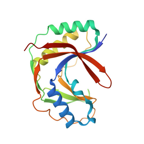
The 2H phosphoesterase family contains enzymes with two His-X-Ser/Thr motifs in the active site. 2H enzymes are found in all kingdoms of life, sharing little sequence identity despite the conserved overall fold and active site. For many 2H enzymes, the physiological function is unknown. Here, we studied the structure of the 2H family member LigT from Escherichia coli both in the apo form and complexed with different active-site ligands, including ATP, 2'-AMP, 3'-AMP, phosphate, and NADP+. Comparisons to the well-characterized vertebrate myelin enzyme 2',3'-cyclic nucleotide 3'-phosphodiesterase (CNPase) highlight specific features of the catalytic cycle and substrate recognition in both enzymes. The role played by the helix α7, unique to CNPases within the 2H family, is apparently taken over by Arg130 in the bacterial enzyme. Other residues and loops lining the active site groove are likely to be important for RNA substrate binding. We visualized conformational changes related to ligand binding, as well as the position of the nucleophilic water molecule. We also present a low-resolution model of E. coli LigT bound to tRNA in solution, and provide a model for RNA binding by LigT, involving flexible loops lining the active site cavity. Taken together, our results both aid in understanding the common features of 2H family enzymes and help highlight the distinct features in the 2H family members, which must result in different reaction mechanisms. Unique aspects in different 2H family members can be observed in ligand recognition and binding, and in the coordination of the nucleophilic water molecule and the reactive phosphate moiety.
Organizational Affiliation:
Faculty of Biochemistry and Molecular Medicine & Biocenter Oulu, University of Oulu, Oulu, Finland.