5L9R

HIF prolyl hydroxylase 2 (PHD2/ EGLN1) in complex with N-oxalylglycine (NOG)


Experimental Data Snapshot

  • Method: X-RAY DIFFRACTION
  • Resolution: 1.81 Å
  • R-Value Free: 0.188 
  • R-Value Work: 0.156 
  • R-Value Observed: 0.158 

wwPDB Validation   3D Report Full Report


This is version 1.3 of the entry. See complete history


Literature

Structural basis for oxygen degradation domain selectivity of the HIF prolyl hydroxylases.

Chowdhury, R.Leung, I.K.Tian, Y.M.Abboud, M.I.Ge, W.Domene, C.Cantrelle, F.X.Landrieu, I.Hardy, A.P.Pugh, C.W.Ratcliffe, P.J.Claridge, T.D.Schofield, C.J.

(2016) Nat Commun 7: 12673-12673

  • DOI: https://doi.org/10.1038/ncomms12673
  • Primary Citation of Related Structures:  
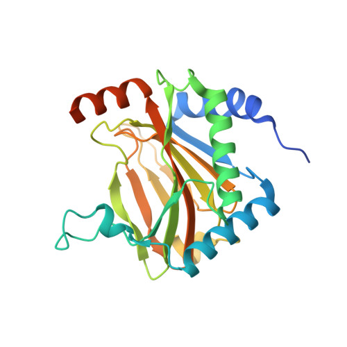
    5L9B, 5L9R, 5L9V, 5LA9, 5LAS, 5LAT, 5LB6, 5LBB, 5LBC, 5LBE, 5LBF

  • PubMed Abstract: 

    The response to hypoxia in animals involves the expression of multiple genes regulated by the αβ-hypoxia-inducible transcription factors (HIFs). The hypoxia-sensing mechanism involves oxygen limited hydroxylation of prolyl residues in the N- and C-terminal oxygen-dependent degradation domains (NODD and CODD) of HIFα isoforms, as catalysed by prolyl hydroxylases (PHD 1-3). Prolyl hydroxylation promotes binding of HIFα to the von Hippel-Lindau protein (VHL)-elongin B/C complex, thus signalling for proteosomal degradation of HIFα. We reveal that certain PHD2 variants linked to familial erythrocytosis and cancer are highly selective for CODD or NODD. Crystalline and solution state studies coupled to kinetic and cellular analyses reveal how wild-type and variant PHDs achieve ODD selectivity via different dynamic interactions involving loop and C-terminal regions. The results inform on how HIF target gene selectivity is achieved and will be of use in developing selective PHD inhibitors.


  • Organizational Affiliation

    Chemistry Research Laboratory, Department of Chemistry, Oxford Centre for Integrative Systems Biology, University of Oxford, Mansfield Road, Oxford OX1 3TA, UK.


Macromolecules
Find similar proteins by:  (by identity cutoff)  |  3D Structure
Entity ID: 1
MoleculeChains Sequence LengthOrganismDetailsImage
Egl nine homolog 1252Homo sapiensMutation(s): 2 
Gene Names: EGLN1C1orf12PNAS-118PNAS-137
EC: 1.14.11.29
UniProt & NIH Common Fund Data Resources
Find proteins for Q9GZT9 (Homo sapiens)
Explore Q9GZT9 
Go to UniProtKB:  Q9GZT9
PHAROS:  Q9GZT9
GTEx:  ENSG00000135766 
Entity Groups  
Sequence Clusters30% Identity50% Identity70% Identity90% Identity95% Identity100% Identity
UniProt GroupQ9GZT9
Sequence Annotations
Expand
  • Reference Sequence
Small Molecules
Ligands 5 Unique
IDChains Name / Formula / InChI Key2D Diagram3D Interactions
PG4
Query on PG4

Download Ideal Coordinates CCD File 
E [auth A]TETRAETHYLENE GLYCOL
C8 H18 O5
UWHCKJMYHZGTIT-UHFFFAOYSA-N
OGA
Query on OGA

Download Ideal Coordinates CCD File 
C [auth A],
D [auth A]
N-OXALYLGLYCINE
C4 H5 N O5
BIMZLRFONYSTPT-UHFFFAOYSA-N
SO4
Query on SO4

Download Ideal Coordinates CCD File 
F [auth A],
G [auth A],
H [auth A]
SULFATE ION
O4 S
QAOWNCQODCNURD-UHFFFAOYSA-L
GOL
Query on GOL

Download Ideal Coordinates CCD File 
I [auth A]GLYCEROL
C3 H8 O3
PEDCQBHIVMGVHV-UHFFFAOYSA-N
MN
Query on MN

Download Ideal Coordinates CCD File 
B [auth A]MANGANESE (II) ION
Mn
WAEMQWOKJMHJLA-UHFFFAOYSA-N
Binding Affinity Annotations 
IDSourceBinding Affinity
OGA BindingDB:  5L9R Ki: 8000 (nM) from 1 assay(s)
Kd: min: 2000, max: 3400 (nM) from 5 assay(s)
IC50: min: 5600, max: 2.60e+4 (nM) from 6 assay(s)
Experimental Data & Validation

Experimental Data

  • Method: X-RAY DIFFRACTION
  • Resolution: 1.81 Å
  • R-Value Free: 0.188 
  • R-Value Work: 0.156 
  • R-Value Observed: 0.158 
  • Space Group: P 41
Unit Cell:
Length ( Å )Angle ( ˚ )
a = 71.233α = 90
b = 71.233β = 90
c = 48.245γ = 90
Software Package:
Software NamePurpose
PHENIXrefinement
HKL-2000data reduction
HKL-2000data scaling
PHASERphasing

Structure Validation

View Full Validation Report


Entry History 

Deposition Data

Revision History  (Full details and data files)

  • Version 1.0: 2016-08-31
    Type: Initial release
  • Version 1.1: 2016-09-07
    Changes: Database references
  • Version 1.2: 2017-11-29
    Changes: Database references
  • Version 1.3: 2024-01-10
    Changes: Data collection, Database references, Derived calculations, Refinement description