5L6N

Disulfated madanin-thrombin complex


Experimental Data Snapshot

  • Method: X-RAY DIFFRACTION
  • Resolution: 1.63 Å
  • R-Value Free: 0.199 
  • R-Value Work: 0.167 
  • R-Value Observed: 0.168 

wwPDB Validation   3D Report Full Report


Ligand Structure Quality Assessment 


This is version 1.3 of the entry. See complete history


Literature

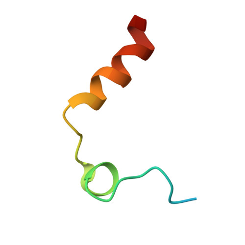
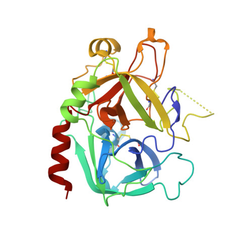
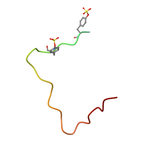
Tyrosine sulfation modulates activity of tick-derived thrombin inhibitors.

Thompson, R.E.Liu, X.Ripoll-Rozada, J.Alonso-Garcia, N.Parker, B.L.Pereira, P.J.B.Payne, R.J.

(2017) Nat Chem 9: 909-917

  • DOI: https://doi.org/10.1038/nchem.2744
  • Primary Citation of Related Structures:  
    5L6N

  • PubMed Abstract: 

    Madanin-1 and chimadanin are two small cysteine-free thrombin inhibitors that facilitate blood feeding in the tick Haemaphysalis longicornis. Here, we report a post-translational modification-tyrosine sulfation-of these two proteins that is critical for potent anti-thrombotic and anticoagulant activity. Inhibitors produced in baculovirus-infected insect cells displayed heterogeneous sulfation of two tyrosine residues within each of the proteins. One-pot ligation-desulfurization chemistry enabled access to homogeneous samples of all possible sulfated variants of the proteins. Tyrosine sulfation of madanin-1 and chimadanin proved crucial for thrombin inhibitory activity, with the doubly sulfated variants three orders of magnitude more potent than the unmodified inhibitors. The three-dimensional structure of madanin-1 in complex with thrombin revealed a unique mode of inhibition, with the sulfated tyrosine residues binding to the basic exosite II of the protease. The importance of tyrosine sulfation within this family of thrombin inhibitors, together with their unique binding mode, paves the way for the development of anti-thrombotic drug leads based on these privileged scaffolds.


  • Organizational Affiliation

    School of Chemistry, The University of Sydney, Sydney, New South Wales 2006, Australia.


Macromolecules
Find similar proteins by:  (by identity cutoff)  |  3D Structure
Entity ID: 1
MoleculeChains Sequence LengthOrganismDetailsImage
ProthrombinA [auth L]36Homo sapiensMutation(s): 0 
EC: 3.4.21.5
UniProt & NIH Common Fund Data Resources
Find proteins for P00734 (Homo sapiens)
Explore P00734 
Go to UniProtKB:  P00734
PHAROS:  P00734
GTEx:  ENSG00000180210 
Entity Groups  
Sequence Clusters30% Identity50% Identity70% Identity90% Identity95% Identity100% Identity
UniProt GroupP00734
Sequence Annotations
Expand
  • Reference Sequence
Find similar proteins by:  (by identity cutoff)  |  3D Structure
Entity ID: 2
MoleculeChains Sequence LengthOrganismDetailsImage
ProthrombinB [auth H]259Homo sapiensMutation(s): 0 
EC: 3.4.21.5
UniProt & NIH Common Fund Data Resources
Find proteins for P00734 (Homo sapiens)
Explore P00734 
Go to UniProtKB:  P00734
PHAROS:  P00734
GTEx:  ENSG00000180210 
Entity Groups  
Sequence Clusters30% Identity50% Identity70% Identity90% Identity95% Identity100% Identity
UniProt GroupP00734
Sequence Annotations
Expand
  • Reference Sequence
Find similar proteins by:  (by identity cutoff)  |  3D Structure
Entity ID: 3
MoleculeChains Sequence LengthOrganismDetailsImage
Thrombin inhibitor madanin 1C [auth I]35Haemaphysalis longicornisMutation(s): 0 
UniProt
Find proteins for Q86FP9 (Haemaphysalis longicornis)
Explore Q86FP9 
Go to UniProtKB:  Q86FP9
Entity Groups  
Sequence Clusters30% Identity50% Identity70% Identity90% Identity95% Identity100% Identity
UniProt GroupQ86FP9
Sequence Annotations
Expand
  • Reference Sequence
Small Molecules
Ligands 2 Unique
IDChains Name / Formula / InChI Key2D Diagram3D Interactions
NAG
Query on NAG

Download Ideal Coordinates CCD File 
D [auth H]2-acetamido-2-deoxy-beta-D-glucopyranose
C8 H15 N O6
OVRNDRQMDRJTHS-FMDGEEDCSA-N
NA
Query on NA

Download Ideal Coordinates CCD File 
E [auth H]SODIUM ION
Na
FKNQFGJONOIPTF-UHFFFAOYSA-N
Modified Residues  1 Unique
IDChains TypeFormula2D DiagramParent
TYS
Query on TYS
C [auth I]L-PEPTIDE LINKINGC9 H11 N O6 STYR
Experimental Data & Validation

Experimental Data

Unit Cell:
Length ( Å )Angle ( ˚ )
a = 44.546α = 90
b = 80.238β = 90
c = 91.369γ = 90
Software Package:
Software NamePurpose
PHENIXrefinement
XDSdata reduction
Aimlessdata scaling
PHASERphasing

Structure Validation

View Full Validation Report


Ligand Structure Quality Assessment 


Entry History 

Deposition Data

Revision History  (Full details and data files)

  • Version 1.0: 2017-03-29
    Type: Initial release
  • Version 1.1: 2017-09-06
    Changes: Database references
  • Version 1.2: 2020-07-29
    Type: Remediation
    Reason: Carbohydrate remediation
    Changes: Data collection, Derived calculations, Structure summary
  • Version 1.3: 2024-01-10
    Changes: Data collection, Database references, Refinement description, Structure summary