5L2W

The X-ray co-crystal structure of human CDK2/CyclinE and Dinaciclib.


Experimental Data Snapshot

  • Method: X-RAY DIFFRACTION
  • Resolution: 2.80 Å
  • R-Value Free: 0.246 
  • R-Value Work: 0.200 
  • R-Value Observed: 0.202 

wwPDB Validation   3D Report Full Report


Ligand Structure Quality Assessment 


This is version 1.2 of the entry. See complete history


Literature

Spectrum and Degree of CDK Drug Interactions Predicts Clinical Performance.

Chen, P.Lee, N.V.Hu, W.Xu, M.Ferre, R.A.Lam, H.Bergqvist, S.Solowiej, J.Diehl, W.He, Y.A.Yu, X.Nagata, A.VanArsdale, T.Murray, B.W.

(2016) Mol Cancer Ther 15: 2273-2281

  • DOI: https://doi.org/10.1158/1535-7163.MCT-16-0300
  • Primary Citation of Related Structures:  
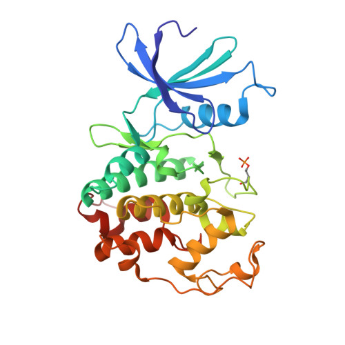
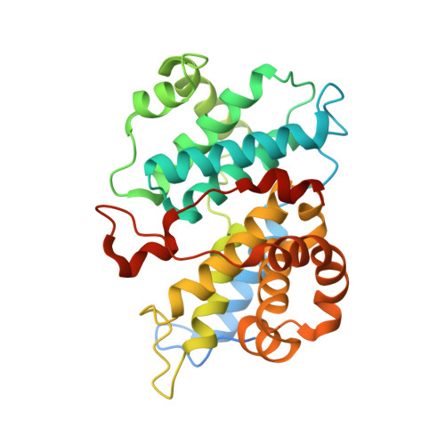
    5L2I, 5L2S, 5L2T, 5L2W

  • PubMed Abstract: 

    Therapeutically targeting aberrant intracellular kinase signaling is attractive from a biological perspective but drug development is often hindered by toxicities and inadequate efficacy. Predicting drug behaviors using cellular and animal models is confounded by redundant kinase activities, a lack of unique substrates, and cell-specific signaling networks. Cyclin-dependent kinase (CDK) drugs exemplify this phenomenon because they are reported to target common processes yet have distinct clinical activities. Tumor cell studies of ATP-competitive CDK drugs (dinaciclib, AG-024322, abemaciclib, palbociclib, ribociclib) indicate similar pharmacology while analyses in untransformed cells illuminates significant differences. To resolve this apparent disconnect, drug behaviors are described at the molecular level. Nonkinase binding studies and kinome interaction analysis (recombinant and endogenous kinases) reveal that proteins outside of the CDK family appear to have little role in dinaciclib/palbociclib/ribociclib pharmacology, may contribute for abemaciclib, and confounds AG-024322 analysis. CDK2 and CDK6 cocrystal structures with the drugs identify the molecular interactions responsible for potency and kinase selectivity. Efficient drug binding to the unique hinge architecture of CDKs enables selectivity toward most of the human kinome. Selectivity between CDK family members is achieved through interactions with nonconserved elements of the ATP-binding pocket. Integrating clinical drug exposures into the analysis predicts that both palbociclib and ribociclib are CDK4/6 inhibitors, abemaciclib inhibits CDK4/6/9, and dinaciclib is a broad-spectrum CDK inhibitor (CDK2/3/4/6/9). Understanding the molecular components of potency and selectivity also facilitates rational design of future generations of kinase-directed drugs. Mol Cancer Ther; 15(10); 2273-81. ©2016 AACR.


  • Organizational Affiliation

    Oncology Medicinal Chemistry, Pfizer Worldwide Research and Development, San Diego, California.


Macromolecules
Find similar proteins by:  (by identity cutoff)  |  3D Structure
Entity ID: 1
MoleculeChains Sequence LengthOrganismDetailsImage
Cyclin-dependent kinase 2299Homo sapiensMutation(s): 0 
Gene Names: CDK2CDKN2
EC: 2.7.11.22
UniProt & NIH Common Fund Data Resources
Find proteins for P24941 (Homo sapiens)
Explore P24941 
Go to UniProtKB:  P24941
PHAROS:  P24941
GTEx:  ENSG00000123374 
Entity Groups  
Sequence Clusters30% Identity50% Identity70% Identity90% Identity95% Identity100% Identity
UniProt GroupP24941
Sequence Annotations
Expand
  • Reference Sequence
Find similar proteins by:  (by identity cutoff)  |  3D Structure
Entity ID: 2
MoleculeChains Sequence LengthOrganismDetailsImage
G1/S-specific cyclin-E1307Homo sapiensMutation(s): 0 
Gene Names: CCNE1CCNE
UniProt & NIH Common Fund Data Resources
Find proteins for P24864 (Homo sapiens)
Explore P24864 
Go to UniProtKB:  P24864
PHAROS:  P24864
GTEx:  ENSG00000105173 
Entity Groups  
Sequence Clusters30% Identity50% Identity70% Identity90% Identity95% Identity100% Identity
UniProt GroupP24864
Sequence Annotations
Expand
  • Reference Sequence
Small Molecules
Ligands 2 Unique
IDChains Name / Formula / InChI Key2D Diagram3D Interactions
1QK
Query on 1QK

Download Ideal Coordinates CCD File 
C [auth A]3-[({3-ethyl-5-[(2S)-2-(2-hydroxyethyl)piperidin-1-yl]pyrazolo[1,5-a]pyrimidin-7-yl}amino)methyl]-1-hydroxypyridinium
C21 H29 N6 O2
WBUFFOKXERTKGU-SFHVURJKSA-N
GOL
Query on GOL

Download Ideal Coordinates CCD File 
D [auth A]GLYCEROL
C3 H8 O3
PEDCQBHIVMGVHV-UHFFFAOYSA-N
Modified Residues  1 Unique
IDChains TypeFormula2D DiagramParent
TPO
Query on TPO
A
L-PEPTIDE LINKINGC4 H10 N O6 PTHR
Experimental Data & Validation

Experimental Data

  • Method: X-RAY DIFFRACTION
  • Resolution: 2.80 Å
  • R-Value Free: 0.246 
  • R-Value Work: 0.200 
  • R-Value Observed: 0.202 
  • Space Group: P 41 21 2
Unit Cell:
Length ( Å )Angle ( ˚ )
a = 100.307α = 90
b = 100.307β = 90
c = 151.857γ = 90
Software Package:
Software NamePurpose
BUSTERrefinement
autoXDSdata processing
BUSTERphasing
Aimlessdata scaling

Structure Validation

View Full Validation Report


Ligand Structure Quality Assessment 


Entry History 

Deposition Data

Revision History  (Full details and data files)

  • Version 1.0: 2016-08-24
    Type: Initial release
  • Version 1.1: 2016-10-19
    Changes: Database references
  • Version 1.2: 2023-10-04
    Changes: Data collection, Database references, Derived calculations, Refinement description