5KYO

Crystal Structure of CYP101J2


Experimental Data Snapshot

  • Method: X-RAY DIFFRACTION
  • Resolution: 1.80 Å
  • R-Value Free: 0.276 
  • R-Value Work: 0.223 
  • R-Value Observed: 0.226 

wwPDB Validation   3D Report Full Report


Ligand Structure Quality Assessment 


This is version 1.2 of the entry. See complete history


Literature

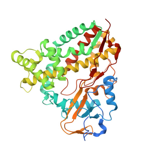
X-ray crystal structure of cytochrome P450 monooxygenase CYP101J2 from Sphingobium yanoikuyae strain B2.

Unterweger, B.Drinkwater, N.Johanesen, P.Lyras, D.Dumsday, G.J.McGowan, S.

(2017) Proteins 85: 945-950

  • DOI: https://doi.org/10.1002/prot.25227
  • Primary Citation of Related Structures:  
    5KYO

  • PubMed Abstract: 

    The cytochrome P450 monooxygenases (P450s) catalyze a vast array of oxygenation reactions that can be useful in biocatalytic applications. CYP101J2 from Sphingobium yanoikuyae is a P450 that catalyzes the hydroxylation of 1,8-cineole. Here we report the crystallization and X-ray structure elucidation of recombinant CYP101J2 to 1.8 Å resolution. The CYP101J2 structure shows the canonical P450-fold and has an open conformation in the absence of substrate. Analysis of the structure revealed that CYP101J2, in the absence of substrate, forms a well-ordered substrate-binding channel that suggests a unique form of substrate guidance in comparison to other bacterial 1,8-cineole-hydroxylating P450 enzymes. Proteins 2017; 85:945-950. © 2016 Wiley Periodicals, Inc.


  • Organizational Affiliation

    Infection and Immunity Program, Biomedicine Discovery Institute, Department of Microbiology, Monash University, Clayton, Victoria, 3800.


Macromolecules
Find similar proteins by:  (by identity cutoff)  |  3D Structure
Entity ID: 1
MoleculeChains Sequence LengthOrganismDetailsImage
CYP101J2
A, B, C, D, E
A, B, C, D, E, F
430Sphingobium yanoikuyaeMutation(s): 0 
UniProt
Find proteins for A0A1C9CIU0 (Sphingobium yanoikuyae)
Explore A0A1C9CIU0 
Go to UniProtKB:  A0A1C9CIU0
Entity Groups  
Sequence Clusters30% Identity50% Identity70% Identity90% Identity95% Identity100% Identity
UniProt GroupA0A1C9CIU0
Sequence Annotations
Expand
  • Reference Sequence
Experimental Data & Validation

Experimental Data

  • Method: X-RAY DIFFRACTION
  • Resolution: 1.80 Å
  • R-Value Free: 0.276 
  • R-Value Work: 0.223 
  • R-Value Observed: 0.226 
  • Space Group: P 1
Unit Cell:
Length ( Å )Angle ( ˚ )
a = 99.426α = 63.46
b = 100.031β = 63.66
c = 99.963γ = 63.67
Software Package:
Software NamePurpose
PHENIXrefinement
iMOSFLMdata reduction
Aimlessdata scaling
PHASERphasing

Structure Validation

View Full Validation Report


Ligand Structure Quality Assessment 


Entry History 

Deposition Data

Revision History  (Full details and data files)

  • Version 1.0: 2017-01-04
    Type: Initial release
  • Version 1.1: 2017-04-26
    Changes: Database references
  • Version 1.2: 2023-10-04
    Changes: Data collection, Database references, Derived calculations, Refinement description