5KYM

Crystal Structure of the 1-acyl-sn-glycerophosphate (LPA) acyltransferase, PlsC, from Thermotoga maritima


Experimental Data Snapshot

  • Method: X-RAY DIFFRACTION
  • Resolution: 2.80 Å
  • R-Value Free: 0.277 
  • R-Value Work: 0.219 
  • R-Value Observed: 0.224 

wwPDB Validation   3D Report Full Report


Ligand Structure Quality Assessment 


This is version 1.3 of the entry. See complete history


Literature

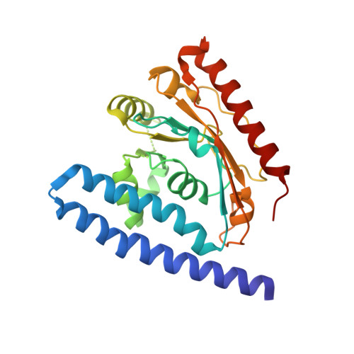
A two-helix motif positions the lysophosphatidic acid acyltransferase active site for catalysis within the membrane bilayer.

Robertson, R.M.Yao, J.Gajewski, S.Kumar, G.Martin, E.W.Rock, C.O.White, S.W.

(2017) Nat Struct Mol Biol 24: 666-671

  • DOI: https://doi.org/10.1038/nsmb.3436
  • Primary Citation of Related Structures:  
    5KYM

  • PubMed Abstract: 

    Phosphatidic acid (PA), the central intermediate in membrane phospholipid synthesis, is generated by two acyltransferases in a pathway conserved in all life forms. The second step in this pathway is catalyzed by 1-acyl-sn-glycerol-3-phosphate acyltransferase, called PlsC in bacteria. Here we present the crystal structure of PlsC from Thermotoga maritima, revealing an unusual hydrophobic/aromatic N-terminal two-helix motif linked to an acyltransferase αβ-domain that contains the catalytic HX 4 D motif. PlsC dictates the acyl chain composition of the 2-position of phospholipids, and the acyl chain selectivity 'ruler' is an appropriately placed and closed hydrophobic tunnel. We confirmed this by site-directed mutagenesis and membrane composition analysis of Escherichia coli cells that expressed mutant PlsC. Molecular dynamics (MD) simulations showed that the two-helix motif represents a novel substructure that firmly anchors the protein to one leaflet of the membrane. This binding mode allows the PlsC active site to acylate lysophospholipids within the membrane bilayer by using soluble acyl donors.


  • Organizational Affiliation

    Department of Structural Biology, St. Jude Children's Research Hospital, Memphis, Tennessee, USA.


Macromolecules
Find similar proteins by:  (by identity cutoff)  |  3D Structure
Entity ID: 1
MoleculeChains Sequence LengthOrganismDetailsImage
1-acyl-sn-glycerol-3-phosphate acyltransferase
A, B
247Thermotoga maritima MSB8Mutation(s): 0 
Gene Names: Tmari_1701
EC: 2.3.1.51
Membrane Entity: Yes 
UniProt
Find proteins for Q9X219 (Thermotoga maritima (strain ATCC 43589 / DSM 3109 / JCM 10099 / NBRC 100826 / MSB8))
Explore Q9X219 
Go to UniProtKB:  Q9X219
Entity Groups  
Sequence Clusters30% Identity50% Identity70% Identity90% Identity95% Identity100% Identity
UniProt GroupQ9X219
Sequence Annotations
Expand
  • Reference Sequence
Small Molecules
Ligands 3 Unique
IDChains Name / Formula / InChI Key2D Diagram3D Interactions
HGX
Query on HGX

Download Ideal Coordinates CCD File 
C [auth A]
D [auth A]
E [auth A]
G [auth B]
H [auth B]
C [auth A],
D [auth A],
E [auth A],
G [auth B],
H [auth B],
I [auth B]
1-HEPTADECANOYL-2-TRIDECANOYL-3-GLYCEROL-PHOSPHONYL CHOLINE
C38 H77 N O8 P
AZEAMUNCULVBPK-PSXMRANNSA-O
LMT
Query on LMT

Download Ideal Coordinates CCD File 
J [auth B]DODECYL-BETA-D-MALTOSIDE
C24 H46 O11
NLEBIOOXCVAHBD-QKMCSOCLSA-N
DD9
Query on DD9

Download Ideal Coordinates CCD File 
F [auth A],
K [auth B]
nonane
C9 H20
BKIMMITUMNQMOS-UHFFFAOYSA-N
Experimental Data & Validation

Experimental Data

  • Method: X-RAY DIFFRACTION
  • Resolution: 2.80 Å
  • R-Value Free: 0.277 
  • R-Value Work: 0.219 
  • R-Value Observed: 0.224 
  • Space Group: I 2 3
Unit Cell:
Length ( Å )Angle ( ˚ )
a = 173.836α = 90
b = 173.836β = 90
c = 173.836γ = 90
Software Package:
Software NamePurpose
REFMACrefinement
XDSdata reduction
XSCALEdata scaling
SHELXCDphasing

Structure Validation

View Full Validation Report


Ligand Structure Quality Assessment 


Entry History 

Deposition Data

Revision History  (Full details and data files)

  • Version 1.0: 2017-07-26
    Type: Initial release
  • Version 1.1: 2017-08-02
    Changes: Database references
  • Version 1.2: 2017-08-16
    Changes: Database references
  • Version 1.3: 2024-03-06
    Changes: Data collection, Database references, Refinement description